- PDB-4jg2: Structure of phage-related protein from Bacillus cereus ATCC 10987 -
+
Open data
ID or keywords:
Loading...
-
Basic information
Entry
Database: PDB / ID: 4jg2
Title
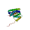
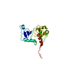
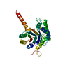
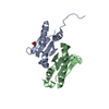
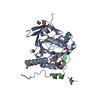
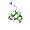
Structure of phage-related protein from Bacillus cereus ATCC 10987
Components
Phage-related protein
Keywords
UNKNOWN FUNCTION / Structural Genomics / PSI-Biology / Midwest Center for Structural Genomics / MCSG / Structures of Mtb Proteins Conferring Susceptibility to Known Mtb Inhibitors / MTBI / phage-related protein
Function / homology
Enterobacter phage Enc34, ssDNA-binding protein / Enterobacter phage Enc34, ssDNA-binding protein / Nucleic acid-binding proteins / OB fold (Dihydrolipoamide Acetyltransferase, E2P) / Nucleic acid-binding, OB-fold / Beta Barrel / Mainly Beta / Phage-related protein
Function and homology information
Biological species
Bacillus cereus (bacteria)
Method
X-RAY DIFFRACTION / SYNCHROTRON / SAD / Resolution: 1.3 Å
Resolution: 1.3→1.32 Å / Redundancy: 3.8 % / Rmerge(I) obs: 0.52 / Mean I/σ(I) obs: 2.5 / % possible all: 97.9
-
Processing
Software
Name
Version
Classification
Blu-Ice
Max
datacollection
PHENIX
modelbuilding
REFMAC
5.7.0032
refinement
XDS
datareduction
XDS
datascaling
PHENIX
phasing
Refinement
Method to determine structure: SAD / Resolution: 1.3→29.89 Å / Cor.coef. Fo:Fc: 0.978 / Cor.coef. Fo:Fc free: 0.968 / SU B: 1.329 / SU ML: 0.026 / Cross valid method: THROUGHOUT / ESU R: 0.046 / ESU R Free: 0.047 / Stereochemistry target values: MAXIMUM LIKELIHOOD / Details: HYDROGENS HAVE BEEN ADDED IN THE RIDING POSITIONS
Rfactor
Num. reflection
% reflection
Selection details
Rfree
0.16349
2127
5.1 %
RANDOM
Rwork
0.12434
-
-
-
obs
0.12631
39942
99.77 %
-
all
-
39942
-
-
Solvent computation
Ion probe radii: 0.8 Å / Shrinkage radii: 0.8 Å / VDW probe radii: 1.2 Å / Solvent model: MASK
Displacement parameters
Biso mean: 15.819 Å2
Baniso -1
Baniso -2
Baniso -3
1-
-0.04 Å2
-0 Å2
-0 Å2
2-
-
0.03 Å2
0 Å2
3-
-
-
0.01 Å2
Refine analyze
Luzzati coordinate error obs: 0.1 Å
Refinement step
Cycle: LAST / Resolution: 1.3→29.89 Å
Protein
Nucleic acid
Ligand
Solvent
Total
Num. atoms
1412
0
0
312
1724
Refine LS restraints
Refine-ID
Type
Dev ideal
Dev ideal target
Number
X-RAY DIFFRACTION
r_bond_refined_d
0.017
0.019
1571
X-RAY DIFFRACTION
r_bond_other_d
0.001
0.02
1436
X-RAY DIFFRACTION
r_angle_refined_deg
1.884
1.945
2154
X-RAY DIFFRACTION
r_angle_other_deg
0.868
3
3318
X-RAY DIFFRACTION
r_dihedral_angle_1_deg
4.949
5
215
X-RAY DIFFRACTION
r_dihedral_angle_2_deg
36.239
25.325
77
X-RAY DIFFRACTION
r_dihedral_angle_3_deg
9.642
15
252
X-RAY DIFFRACTION
r_dihedral_angle_4_deg
15.808
15
9
X-RAY DIFFRACTION
r_chiral_restr
0.13
0.2
231
X-RAY DIFFRACTION
r_gen_planes_refined
0.01
0.021
1940
X-RAY DIFFRACTION
r_gen_planes_other
0.002
0.02
369
X-RAY DIFFRACTION
r_nbd_refined
X-RAY DIFFRACTION
r_nbd_other
X-RAY DIFFRACTION
r_nbtor_refined
X-RAY DIFFRACTION
r_nbtor_other
X-RAY DIFFRACTION
r_xyhbond_nbd_refined
X-RAY DIFFRACTION
r_xyhbond_nbd_other
X-RAY DIFFRACTION
r_metal_ion_refined
X-RAY DIFFRACTION
r_metal_ion_other
X-RAY DIFFRACTION
r_symmetry_vdw_refined
X-RAY DIFFRACTION
r_symmetry_vdw_other
X-RAY DIFFRACTION
r_symmetry_hbond_refined
X-RAY DIFFRACTION
r_symmetry_hbond_other
X-RAY DIFFRACTION
r_symmetry_metal_ion_refined
X-RAY DIFFRACTION
r_symmetry_metal_ion_other
X-RAY DIFFRACTION
r_mcbond_it
1.998
0.967
821
X-RAY DIFFRACTION
r_mcbond_other
1.982
0.963
820
X-RAY DIFFRACTION
r_mcangle_it
2.312
1.458
1049
X-RAY DIFFRACTION
r_scbond_it
4.116
1.291
750
X-RAY DIFFRACTION
r_scangle_it
X-RAY DIFFRACTION
r_rigid_bond_restr
4.95
3
3007
X-RAY DIFFRACTION
r_sphericity_free
35.811
5
74
X-RAY DIFFRACTION
r_sphericity_bonded
11.395
5
3213
LS refinement shell
Resolution: 1.3→1.334 Å / Total num. of bins used: 20
Rfactor
Num. reflection
% reflection
Rfree
0.265
155
-
Rwork
0.219
2850
-
obs
-
-
98.11 %
Refinement TLS params.
Method: refined / Refine-ID: X-RAY DIFFRACTION
ID
L11 (°2)
L12 (°2)
L13 (°2)
L22 (°2)
L23 (°2)
L33 (°2)
S11 (Å °)
S12 (Å °)
S13 (Å °)
S21 (Å °)
S22 (Å °)
S23 (Å °)
S31 (Å °)
S32 (Å °)
S33 (Å °)
T11 (Å2)
T12 (Å2)
T13 (Å2)
T22 (Å2)
T23 (Å2)
T33 (Å2)
Origin x (Å)
Origin y (Å)
Origin z (Å)
1
0.6853
-0.1107
-0.0788
0.9785
0.1333
0.476
0.011
-0.0007
0.0268
0.0025
-0.0168
-0.0003
-0.0396
0.0035
0.0059
0.0125
-0.0015
0.0021
0.0179
0.0001
0.0019
14.6483
-2.3089
0.4202
2
1.0961
-0.5988
-0.459
0.6055
0.4724
0.7124
0.083
0.1074
0.1244
-0.0974
-0.0219
-0.1045
-0.0943
0.0193
-0.061
0.032
-0.0026
0.026
0.0265
0.0165
0.0437
24.1835
5.4713
-8.5197
3
0.302
0.1135
-0.0236
0.2007
-0.0195
0.1504
0.0305
-0.02
-0.0008
0.0183
-0.0246
-0.0053
-0.0176
0.0073
-0.0059
0.0146
0.0037
0.005
0.0121
0.0023
0.0072
11.1558
-6.858
-0.0435
Refinement TLS group
ID
Refine-ID
Refine TLS-ID
Auth asym-ID
Auth seq-ID
1
X-RAY DIFFRACTION
1
A
4 - 49
2
X-RAY DIFFRACTION
2
A
50 - 90
3
X-RAY DIFFRACTION
3
A
91 - 176
+
About Yorodumi
-
News
-
Feb 9, 2022. New format data for meta-information of EMDB entries
New format data for meta-information of EMDB entries
Version 3 of the EMDB header file is now the official format.
The previous official version 1.9 will be removed from the archive.
In the structure databanks used in Yorodumi, some data are registered as the other names, "COVID-19 virus" and "2019-nCoV". Here are the details of the virus and the list of structure data.
Jan 31, 2019. EMDB accession codes are about to change! (news from PDBe EMDB page)
EMDB accession codes are about to change! (news from PDBe EMDB page)
The allocation of 4 digits for EMDB accession codes will soon come to an end. Whilst these codes will remain in use, new EMDB accession codes will include an additional digit and will expand incrementally as the available range of codes is exhausted. The current 4-digit format prefixed with “EMD-” (i.e. EMD-XXXX) will advance to a 5-digit format (i.e. EMD-XXXXX), and so on. It is currently estimated that the 4-digit codes will be depleted around Spring 2019, at which point the 5-digit format will come into force.
The EM Navigator/Yorodumi systems omit the EMD- prefix.
Related info.:Q: What is EMD? / ID/Accession-code notation in Yorodumi/EM Navigator
Yorodumi is a browser for structure data from EMDB, PDB, SASBDB, etc.
This page is also the successor to EM Navigator detail page, and also detail information page/front-end page for Omokage search.
The word "yorodu" (or yorozu) is an old Japanese word meaning "ten thousand". "mi" (miru) is to see.
Related info.:EMDB / PDB / SASBDB / Comparison of 3 databanks / Yorodumi Search / Aug 31, 2016. New EM Navigator & Yorodumi / Yorodumi Papers / Jmol/JSmol / Function and homology information / Changes in new EM Navigator and Yorodumi
 Movie
Movie Controller

 Yorodumi
 Open data
 Basic information
 Components
 Keywords
 Function and homology information
 X-RAY DIFFRACTION /
  Authors
 Citation
 Structure visualization
 Downloads & links
 Other downloads
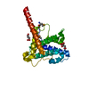

 PDBj
PDBj Assembly
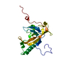
 Mass: 18.015 Da / Num. of mol.: 312 / Source method: isolated from a natural source / Formula: H2O
 Sample preparation
 / Beamline: 21-ID-G / Wavelength: 0.97856 Å
 Processing