 Movie
Movie Controller


+ Open data

- Basic information
| Entry | Database: PDB / ID: 1n0w | ||||||
|---|---|---|---|---|---|---|---|
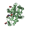
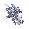
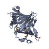
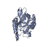
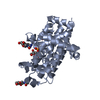
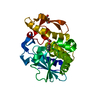
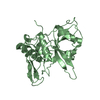
| Title | Crystal structure of a RAD51-BRCA2 BRC repeat complex | ||||||
 Components Components |
| ||||||
 Keywords Keywords | GENE REGULATION/ANTITUMOR PROTEIN / DNA repair / homologous recombination / breast cancer susceptibility / RecA-like ATPase / protein complex / GENE REGULATION-ANTITUMOR PROTEIN COMPLEX | ||||||
| Function / homology |  Function and homology information Function and homology informationBRCA2-MAGE-D1 complex / negative regulation of mammary gland epithelial cell proliferation / nuclear ubiquitin ligase complex / presynaptic intermediate filament cytoskeleton / response to glucoside / establishment of protein localization to telomere / mitotic recombination-dependent replication fork processing / DNA recombinase assembly / cellular response to cisplatin / chromosome organization involved in meiotic cell cycle ...BRCA2-MAGE-D1 complex / negative regulation of mammary gland epithelial cell proliferation / nuclear ubiquitin ligase complex / presynaptic intermediate filament cytoskeleton / response to glucoside / establishment of protein localization to telomere / mitotic recombination-dependent replication fork processing / DNA recombinase assembly / cellular response to cisplatin / chromosome organization involved in meiotic cell cycle / telomere maintenance via telomere lengthening / double-strand break repair involved in meiotic recombination / histone H4 acetyltransferase activity / oocyte maturation / DNA strand invasion / histone H3 acetyltransferase activity / mitotic recombination / cellular response to camptothecin / lateral element / Impaired BRCA2 translocation to the nucleus / Impaired BRCA2 binding to SEM1 (DSS1) / DNA strand exchange activity / gamma-tubulin binding / HDR through MMEJ (alt-NHEJ) / Impaired BRCA2 binding to PALB2 / regulation of DNA damage checkpoint / DNA repair complex / telomere maintenance via recombination / inner cell mass cell proliferation / response to UV-C / reciprocal meiotic recombination / single-stranded DNA helicase activity / ATP-dependent DNA damage sensor activity / regulation of double-strand break repair via homologous recombination / HDR through Single Strand Annealing (SSA) / hematopoietic stem cell proliferation / nuclear chromosome / Homologous DNA Pairing and Strand Exchange / Defective homologous recombination repair (HRR) due to BRCA1 loss of function / Defective HDR through Homologous Recombination Repair (HRR) due to PALB2 loss of BRCA1 binding function / Defective HDR through Homologous Recombination Repair (HRR) due to PALB2 loss of BRCA2/RAD51/RAD51C binding function / Resolution of D-loop Structures through Synthesis-Dependent Strand Annealing (SDSA) / female gonad development / Resolution of D-loop Structures through Holliday Junction Intermediates / Transcriptional Regulation by E2F6 / centrosome duplication / Impaired BRCA2 binding to RAD51 / male meiosis I / replication fork processing / intrinsic apoptotic signaling pathway in response to DNA damage by p53 class mediator / Presynaptic phase of homologous DNA pairing and strand exchange / response to X-ray / ATP-dependent activity, acting on DNA / interstrand cross-link repair / positive regulation of mitotic cell cycle / condensed chromosome / DNA polymerase binding / secretory granule / condensed nuclear chromosome / response to gamma radiation / regulation of cytokinesis / cellular response to ionizing radiation / DNA damage response, signal transduction by p53 class mediator / nucleotide-excision repair / brain development / meiotic cell cycle / cellular response to gamma radiation / protein-DNA complex / PML body / double-strand break repair via homologous recombination / Meiotic recombination / Hydrolases; Acting on acid anhydrides; Acting on acid anhydrides to facilitate cellular and subcellular movement / response to toxic substance / HDR through Homologous Recombination (HRR) / cellular senescence / double-strand break repair / single-stranded DNA binding / site of double-strand break / protease binding / double-stranded DNA binding / DNA recombination / spermatogenesis / chromosome, telomeric region / response to xenobiotic stimulus / mitochondrial matrix / hydrolase activity / DNA repair / chromatin binding / centrosome / regulation of DNA-templated transcription / DNA damage response / nucleolus / positive regulation of DNA-templated transcription / chromatin / perinuclear region of cytoplasm / enzyme binding / protein-containing complex / mitochondrion / nucleoplasm / ATP binding Similarity search - Function | ||||||
| Biological species |  Homo sapiens (human) Homo sapiens (human) | ||||||
| Method |  X-RAY DIFFRACTION / SYNCHROTRON / MAD / Resolution: 1.7 Å X-RAY DIFFRACTION / SYNCHROTRON / MAD / Resolution: 1.7 Å | ||||||
 Authors Authors | Pellegrini, L. / Yu, D.S. / Lo, T. / Anand, S. / Lee, M. / Blundell, T.L. / Venkitaraman, A.R. | ||||||
 Citation Citation | Journal: Nature / Year: 2002 Title: Insights into DNA recombination from the structure of a RAD51-BRCA2 complex Authors: Pellegrini, L. / Yu, D.S. / Lo, T. / Anand, S. / Lee, M. / Blundell, T.L. / Venkitaraman, A.R. | ||||||
| History |
| ||||||
| Remark 999 | SEQUENCE All chains in the entry are part of an engineered, chimeric protein construct. In the ...SEQUENCE All chains in the entry are part of an engineered, chimeric protein construct. In the construct, the amino-to-carboxyl order of the chains is: C-B-L-A. Chain C is a GSMG sequence that comes from the expression vector, B is the BRCA2 BRC repeat, L is the TGSTGSTGSTGSMG sequence linking chains B and A, and chain A is the RAD51 ATpase domain. Chain C appears as a separate entity because the two initial residues of chain B, 1517-8, are not visible in the density map. Likewise, the linker is not visible in the density map, with the exception of the first three residues:TGS. |
- Structure visualization
| Structure viewer | Molecule: MolmilJmol/JSmol |
|---|
- Downloads & links
-Download
| PDBx/mmCIF format | 1n0w.cif.gz | 72.1 KB | Display | PDBx/mmCIF format |
|---|---|---|---|---|
| PDB format | pdb1n0w.ent.gz | 52.2 KB | Display | PDB format |
| PDBx/mmJSON format | 1n0w.json.gz | Tree view | PDBx/mmJSON format | |
| Others |  Other downloads Other downloads |
-Validation report
| Arichive directory | https://data.pdbj.org/pub/pdb/validation_reports/n0/1n0wftp://data.pdbj.org/pub/pdb/validation_reports/n0/1n0w | HTTPS FTP |
|---|
-Related structure data
| Similar structure data |
|---|


-Links
 PDBj
PDBj


- Assembly
| Deposited unit | 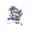
| ||||||||
|---|---|---|---|---|---|---|---|---|---|
| 1 | 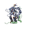
| ||||||||
| Unit cell |
| ||||||||
| Details | The asymmetric unit contains one biological unit, constituted by one chain A, one chain B and one chain C |
-Components
-Protein , 1 types, 1 molecules A
| #1: Protein | Mass: 26855.346 Da / Num. of mol.: 1 / Fragment: ATPase domain Source method: isolated from a genetically manipulated source Source: (gene. exp.) Homo sapiens (human) / Gene: RAD51 / Plasmid: pGAT3 / Species (production host): Escherichia coli / Production host:  |
|---|
-Protein/peptide , 3 types, 3 molecules BLC
| #2: Protein/peptide | Mass: 3966.598 Da / Num. of mol.: 1 / Fragment: BRC repeat type 4 Source method: isolated from a genetically manipulated source Source: (gene. exp.) Homo sapiens (human) / Gene: BRCA2 / Plasmid: pGAT3 / Species (production host): Escherichia coli / Production host: |
|---|---|
| #3: Protein/peptide | Mass: 1234.091 Da / Num. of mol.: 1 Source method: isolated from a genetically manipulated source Details: this peptide links the BRCA2 to the RAD51 / Plasmid: pGAT3 / Species (production host): Escherichia coli / Production host: |
| #4: Protein/peptide | Mass: 397.287 Da / Num. of mol.: 1 Source method: isolated from a genetically manipulated source Details: this peptide comes from the expression vector and is linked to the N-terminus of BRCA2 Plasmid: pGAT3 / Species (production host): Escherichia coli / Production host: |
-Non-polymers , 4 types, 254 molecules 


| #5: Chemical | ChemComp-MG /  Mass: 24.305 Da / Num. of mol.: 1 / Source method: obtained synthetically / Formula: Mg Mass: 24.305 Da / Num. of mol.: 1 / Source method: obtained synthetically / Formula: Mg | ||
|---|---|---|---|
| #6: Chemical | ChemComp-CL /  Mass: 35.453 Da / Num. of mol.: 1 / Source method: obtained synthetically / Formula: Cl Mass: 35.453 Da / Num. of mol.: 1 / Source method: obtained synthetically / Formula: Cl | ||
| #7: Chemical | ChemComp-EDO /  Mass: 62.068 Da / Num. of mol.: 5 / Source method: obtained synthetically / Formula: C2H6O2 Mass: 62.068 Da / Num. of mol.: 5 / Source method: obtained synthetically / Formula: C2H6O2#8: Water | ChemComp-HOH / | Mass: 18.015 Da / Num. of mol.: 247 / Source method: isolated from a natural source / Formula: H2O |
-Details
| Has protein modification | Y |
|---|
-Experimental details
-Experiment
| Experiment | Method: X-RAY DIFFRACTION / Number of used crystals: 1 |
|---|
- Sample preparation
| Crystal | Density Matthews: 2.01 Å3/Da / Density % sol: 38.95 % / Description: Freidel pairs were used. |
|---|---|
| Crystal grow | Temperature: 291 K / Method: vapor diffusion, hanging drop / pH: 7.2 Details: ETHYLENE GLYCOL, pH 7.2, VAPOR DIFFUSION, HANGING DROP, temperature 291K |
| Crystal grow | *PLUS Temperature: 18 ℃ |
| Components of the solutions | *PLUS Conc.: 25 % / Common name: ethylene glycol |
-Data collection
| Diffraction | Mean temperature: 100 K | |||||||||
|---|---|---|---|---|---|---|---|---|---|---|
| Diffraction source | Source: SYNCHROTRON / Site: ESRF  / Beamline: ID29 / Wavelength: 0.9792, 0.90831 / Beamline: ID29 / Wavelength: 0.9792, 0.90831 | |||||||||
| Detector | Type: ADSC QUANTUM 4 / Detector: CCD / Date: Apr 7, 2002 | |||||||||
| Radiation | Monochromator: channel - cut Si monochromator + cylindrical grazing incidence mirror Protocol: MAD / Monochromatic (M) / Laue (L): M / Scattering type: x-ray | |||||||||
| Radiation wavelength |
| |||||||||
| Reflection | Resolution: 1.7→24.85 Å / Num. obs: 55746 / % possible obs: 99.9 % / Observed criterion σ(F): 0 / Observed criterion σ(I): 0 / Redundancy: 7 % / Biso Wilson estimate: 16.9 Å2 / Rmerge(I) obs: 0.077 / Rsym value: 0.077 / Net I/σ(I): 23.5 | |||||||||
| Reflection shell | Resolution: 1.7→1.73 Å / Rmerge(I) obs: 0.321 / Mean I/σ(I) obs: 6.5 / Rsym value: 0.321 / % possible all: 99.9 | |||||||||
| Reflection | *PLUS Highest resolution: 1.7 Å / Num. obs: 29143 / Num. measured all: 204230 | |||||||||
| Reflection shell | *PLUS % possible obs: 99.9 % |
- Processing
| Software |
| ||||||||||||||||||||
|---|---|---|---|---|---|---|---|---|---|---|---|---|---|---|---|---|---|---|---|---|---|
| Refinement | Method to determine structure: MAD / Resolution: 1.7→24.85 Å / Data cutoff high absF: 0 / Cross valid method: THROUGHOUT / σ(F): 0 / σ(I): 0 / Stereochemistry target values: Engh & Huber Details: The structure was refined against the peak Se K edge wavelength dataset. Selenomethionine residues (residue name MSE) were used in the refinement, and are present in the deposited coordinate ...Details: The structure was refined against the peak Se K edge wavelength dataset. Selenomethionine residues (residue name MSE) were used in the refinement, and are present in the deposited coordinate file. Freidel pairs were used.
| ||||||||||||||||||||
| Displacement parameters | Biso mean: 21.1 Å2 | ||||||||||||||||||||
| Refinement step | Cycle: LAST / Resolution: 1.7→24.85 Å
| ||||||||||||||||||||
| Refine LS restraints |
| ||||||||||||||||||||
| Refinement | *PLUS Highest resolution: 1.7 Å / Lowest resolution: 24.8 Å | ||||||||||||||||||||
| Solvent computation | *PLUS | ||||||||||||||||||||
| Displacement parameters | *PLUS | ||||||||||||||||||||
| Refine LS restraints | *PLUS Type: c_angle_deg / Dev ideal: 1.229 |
