 Movie
Movie Controller


+ Open data

- Basic information
| Entry | Database: PDB / ID: 3zn4 | ||||||
|---|---|---|---|---|---|---|---|
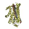
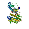
| Title | VP16, a capsid protein of bacteriophage P23-77 (VP16-type-2) | ||||||
 Components Components | VP16 | ||||||
 Keywords Keywords | VIRAL PROTEIN | ||||||
| Function / homology | Jelly Rolls - #1180 / : / Major capsid protein VP16-like / Jelly Rolls / Sandwich / Mainly Beta / identical protein binding / CITRIC ACID / VP16 Function and homology information Function and homology information | ||||||
| Biological species |   THERMUS PHAGE P23-77 (virus) THERMUS PHAGE P23-77 (virus) | ||||||
| Method |  X-RAY DIFFRACTION / SYNCHROTRON / MOLECULAR REPLACEMENT / Resolution: 1.26 Å X-RAY DIFFRACTION / SYNCHROTRON / MOLECULAR REPLACEMENT / Resolution: 1.26 Å | ||||||
 Authors Authors | Rissanen, I. / Grimes, J.M. / Pawlowski, A. / Mantynen, S. / Harlos, K. / Bamford, J.K.H. / Stuart, D.I. | ||||||
 Citation Citation | Journal: Structure / Year: 2013 Title: Bacteriophage P23-77 Capsid Protein Structures Reveal the Archetype of an Ancient Branch from a Major Virus Lineage. Authors: Rissanen, I. / Grimes, J.M. / Pawlowski, A. / Mantynen, S. / Harlos, K. / Bamford, J.K.H. / Stuart, D.I. | ||||||
| History |
|
- Structure visualization
| Structure viewer | Molecule: MolmilJmol/JSmol |
|---|
- Downloads & links
-Download
| PDBx/mmCIF format | 3zn4.cif.gz | 81.8 KB | Display | PDBx/mmCIF format |
|---|---|---|---|---|
| PDB format | pdb3zn4.ent.gz | 60.8 KB | Display | PDB format |
| PDBx/mmJSON format | 3zn4.json.gz | Tree view | PDBx/mmJSON format | |
| Others |  Other downloads Other downloads |
-Validation report
| Arichive directory | https://data.pdbj.org/pub/pdb/validation_reports/zn/3zn4ftp://data.pdbj.org/pub/pdb/validation_reports/zn/3zn4 | HTTPS FTP |
|---|
-Related structure data
| Related structure data | 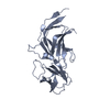 3zmnC 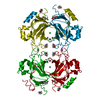 3zmoSC 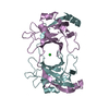 3zn5C 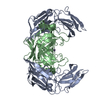 3zn6C C: citing same article ( S: Starting model for refinement |
|---|---|
| Similar structure data |


-Links
 PDBj
PDBj- Assembly
| Deposited unit | 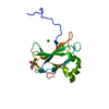
| ||||||||||||||||||
|---|---|---|---|---|---|---|---|---|---|---|---|---|---|---|---|---|---|---|---|
| 1 | 
| ||||||||||||||||||
| Unit cell |
| ||||||||||||||||||
| Components on special symmetry positions |
|

-Components
| #1: Protein | Mass: 19127.773 Da / Num. of mol.: 1 Source method: isolated from a genetically manipulated source Details: SUBUNIT OF A STRAND-SWAPPED HOMODIMER / Source: (gene. exp.) THERMUS PHAGE P23-77 (virus) / Plasmid: PIR2 (ORF16/PET22B) / Production host:  |
|---|---|
| #2: Chemical | ChemComp-CIT /   Mass: 192.124 Da / Num. of mol.: 1 / Source method: obtained synthetically / Formula: C6H8O7 Mass: 192.124 Da / Num. of mol.: 1 / Source method: obtained synthetically / Formula: C6H8O7 |
| #3: Chemical | ChemComp-CL /   Mass: 35.453 Da / Num. of mol.: 1 / Source method: obtained synthetically / Formula: Cl Mass: 35.453 Da / Num. of mol.: 1 / Source method: obtained synthetically / Formula: Cl |
| #4: Water | ChemComp-HOH /  Mass: 18.015 Da / Num. of mol.: 255 / Source method: isolated from a natural source / Formula: H2O Mass: 18.015 Da / Num. of mol.: 255 / Source method: isolated from a natural source / Formula: H2O |
-Experimental details
-Experiment
| Experiment | Method: X-RAY DIFFRACTION / Number of used crystals: 1 |
|---|
- Sample preparation
| Crystal | Density Matthews: 2.15 Å3/Da / Density % sol: 39 % / Description: NONE |
|---|---|
| Crystal grow | Method: vapor diffusion, sitting drop / pH: 4 Details: SITTING DROP VAPOUR DIFFUSION SYSTEM IN A 96-WELL PLATE, 200 NL OF PROTEIN (2-3 MG/ML IN 20 MM TRIS-BUFFER PH 7.4) MIXED WITH 200 NL SOLUTION CONSISTING 20%(W/V) PEG6000 AND 0.1 M CITRATE PH 4. |
-Data collection
| Diffraction | Mean temperature: 100 K |
|---|---|
| Diffraction source | Source: SYNCHROTRON / Site: Diamond  / Beamline: I04 / Wavelength: 1 / Beamline: I04 / Wavelength: 1 |
| Detector | Type: ADSC CCD / Detector: CCD / Date: Jun 26, 2010 |
| Radiation | Protocol: SINGLE WAVELENGTH / Monochromatic (M) / Laue (L): M / Scattering type: x-ray |
| Radiation wavelength | Wavelength: 1 Å / Relative weight: 1 |
| Reflection | Resolution: 1.26→34.3 Å / Num. obs: 37146 / % possible obs: 85.8 % / Redundancy: 7.5 % / Biso Wilson estimate: 10.67 Å2 / Rmerge(I) obs: 0.05 / Net I/σ(I): 23.1 |
| Reflection shell | Resolution: 1.26→1.3 Å / Redundancy: 6.5 % / Rmerge(I) obs: 0.63 / Mean I/σ(I) obs: 2.6 / % possible all: 41.4 |
- Processing
| Software |
| |||||||||||||||||||||||||||||||||||||||||||||||||||||||||||||||||||||||||||||||||||||||||||||||||||||||||||||||||||||||||||||||||||||||||||||||||||||||||||||||||||||||||||||||
|---|---|---|---|---|---|---|---|---|---|---|---|---|---|---|---|---|---|---|---|---|---|---|---|---|---|---|---|---|---|---|---|---|---|---|---|---|---|---|---|---|---|---|---|---|---|---|---|---|---|---|---|---|---|---|---|---|---|---|---|---|---|---|---|---|---|---|---|---|---|---|---|---|---|---|---|---|---|---|---|---|---|---|---|---|---|---|---|---|---|---|---|---|---|---|---|---|---|---|---|---|---|---|---|---|---|---|---|---|---|---|---|---|---|---|---|---|---|---|---|---|---|---|---|---|---|---|---|---|---|---|---|---|---|---|---|---|---|---|---|---|---|---|---|---|---|---|---|---|---|---|---|---|---|---|---|---|---|---|---|---|---|---|---|---|---|---|---|---|---|---|---|---|---|---|---|---|
| Refinement | Method to determine structure: MOLECULAR REPLACEMENT Starting model: PDB ENTRY 3ZMO Resolution: 1.26→17.79 Å / Cor.coef. Fo:Fc: 0.9655 / Cor.coef. Fo:Fc free: 0.9548 / SU R Cruickshank DPI: 0.046 / Cross valid method: THROUGHOUT / σ(F): 0 / SU R Blow DPI: 0.049 / SU Rfree Blow DPI: 0.052 / SU Rfree Cruickshank DPI: 0.049 Details: IDEAL-DIST CONTACT TERM CONTACT SETUP. ALL ATOMS HAVE CCP4 ATOM TYPE FROM LIBRARY. RESIDUES 1-18 AND 168-173 ARE ALTERNATE CONFORMATIONS WERE MODELLED.
| |||||||||||||||||||||||||||||||||||||||||||||||||||||||||||||||||||||||||||||||||||||||||||||||||||||||||||||||||||||||||||||||||||||||||||||||||||||||||||||||||||||||||||||||
| Displacement parameters | Biso mean: 16.06 Å2
| |||||||||||||||||||||||||||||||||||||||||||||||||||||||||||||||||||||||||||||||||||||||||||||||||||||||||||||||||||||||||||||||||||||||||||||||||||||||||||||||||||||||||||||||
| Refine analyze | Luzzati coordinate error obs: 0.13 Å | |||||||||||||||||||||||||||||||||||||||||||||||||||||||||||||||||||||||||||||||||||||||||||||||||||||||||||||||||||||||||||||||||||||||||||||||||||||||||||||||||||||||||||||||
| Refinement step | Cycle: LAST / Resolution: 1.26→17.79 Å
| |||||||||||||||||||||||||||||||||||||||||||||||||||||||||||||||||||||||||||||||||||||||||||||||||||||||||||||||||||||||||||||||||||||||||||||||||||||||||||||||||||||||||||||||
| Refine LS restraints |
| |||||||||||||||||||||||||||||||||||||||||||||||||||||||||||||||||||||||||||||||||||||||||||||||||||||||||||||||||||||||||||||||||||||||||||||||||||||||||||||||||||||||||||||||
| LS refinement shell | Resolution: 1.26→1.29 Å / Total num. of bins used: 19
| |||||||||||||||||||||||||||||||||||||||||||||||||||||||||||||||||||||||||||||||||||||||||||||||||||||||||||||||||||||||||||||||||||||||||||||||||||||||||||||||||||||||||||||||
| Refinement TLS params. | Method: refined / Refine-ID: X-RAY DIFFRACTION
| |||||||||||||||||||||||||||||||||||||||||||||||||||||||||||||||||||||||||||||||||||||||||||||||||||||||||||||||||||||||||||||||||||||||||||||||||||||||||||||||||||||||||||||||
| Refinement TLS group |
|
