[English] 日本語
Yorodumi
- PDB-6pii: The evolving story of AtzT, a periplasmic binding protein -

+
Open data


ID or keywords:

Loading...

-
Basic information

Entry
Database: PDB / ID: 6pii
TitleThe evolving story of AtzT, a periplasmic binding protein
ComponentsAtrazine periplasmic binding protein
KeywordsTRANSPORT PROTEIN / SAD phasing / periplasmic binding protein / evolution from purine binding to atrazine binding
Function / homology
Function and homology information


metal ion binding / plasma membrane
Similarity search - Function
ABC transporter substrate-binding protein PnrA-like / ABC transporter substrate-binding protein PnrA-like / Response regulator / Periplasmic binding protein-like I / Rossmann fold / 3-Layer(aba) Sandwich / Alpha Beta
Similarity search - Domain/homology
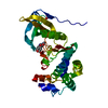
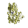
ETHYL MERCURY ION / GUANINE / PHOSPHATE ION / ABC transporter substrate-binding protein PnrA-like domain-containing protein
Similarity search - Component
Biological species
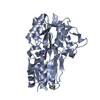
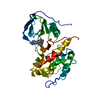
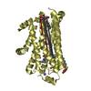
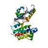
Pseudomonas sp. (bacteria)
MethodX-RAY DIFFRACTION / SYNCHROTRON / SAD / Resolution: 1.87 Å
AuthorsPeat, T.S. / Newman, J. / Scott, C. / Esquirol, L. / Dennis, M. / Nebl, T.
CitationJournal: Acta Crystallogr D Struct Biol / Year: 2019
Title: The evolving story of AtzT, a periplasmic binding protein.
Authors: Dennis, M.L. / Esquirol, L. / Nebl, T. / Newman, J. / Scott, C. / Peat, T.S.
History
DepositionJun 26, 2019Deposition site: RCSB / Processing site: RCSB
Revision 1.0Nov 13, 2019Provider: repository / Type: Initial release
Revision 1.1Dec 25, 2019Group: Database references / Category: citation / citation_author
Item: _citation.country / _citation.journal_abbrev ..._citation.country / _citation.journal_abbrev / _citation.journal_id_ASTM / _citation.journal_id_ISSN / _citation.page_last / _citation.pdbx_database_id_PubMed / _citation.title / _citation_author.identifier_ORCID / _citation_author.name
Revision 1.2Mar 13, 2024Group: Data collection / Database references ...Data collection / Database references / Derived calculations / Refinement description
Category: chem_comp_atom / chem_comp_bond ...chem_comp_atom / chem_comp_bond / database_2 / struct_conn / struct_ncs_dom_lim
Item: _database_2.pdbx_DOI / _database_2.pdbx_database_accession ..._database_2.pdbx_DOI / _database_2.pdbx_database_accession / _struct_conn.pdbx_dist_value / _struct_conn.ptnr1_auth_asym_id / _struct_conn.ptnr1_auth_comp_id / _struct_conn.ptnr1_auth_seq_id / _struct_conn.ptnr1_label_asym_id / _struct_conn.ptnr1_label_atom_id / _struct_conn.ptnr1_label_comp_id / _struct_conn.ptnr1_label_seq_id / _struct_conn.ptnr2_auth_asym_id / _struct_conn.ptnr2_auth_comp_id / _struct_conn.ptnr2_auth_seq_id / _struct_conn.ptnr2_label_asym_id / _struct_conn.ptnr2_label_atom_id / _struct_conn.ptnr2_label_comp_id / _struct_ncs_dom_lim.beg_auth_comp_id / _struct_ncs_dom_lim.beg_label_asym_id / _struct_ncs_dom_lim.beg_label_comp_id / _struct_ncs_dom_lim.beg_label_seq_id / _struct_ncs_dom_lim.end_auth_comp_id / _struct_ncs_dom_lim.end_label_asym_id / _struct_ncs_dom_lim.end_label_comp_id / _struct_ncs_dom_lim.end_label_seq_id

-
Structure visualization

Structure viewerMolecule:
MolmilJmol/JSmol

Downloads & links

-
Assembly

Deposited unit
A: Atrazine periplasmic binding protein
B: Atrazine periplasmic binding protein
C: Atrazine periplasmic binding protein
D: Atrazine periplasmic binding protein
hetero molecules


Theoretical massNumber of molelcules
Total (without water)154,92015
Polymers153,1674
Non-polymers1,75311
Water11,674648
1
A: Atrazine periplasmic binding protein
hetero molecules


Theoretical massNumber of molelcules
Total (without water)38,7674
Polymers38,2921
Non-polymers4763
Water181
TypeNameSymmetry operationNumber
identity operation1_555x,y,z1
MethodPISA
2
B: Atrazine periplasmic binding protein
hetero molecules


Theoretical massNumber of molelcules
Total (without water)38,7674
Polymers38,2921
Non-polymers4763
Water181
TypeNameSymmetry operationNumber
identity operation1_555x,y,z1
MethodPISA
3
C: Atrazine periplasmic binding protein
hetero molecules


Theoretical massNumber of molelcules
Total (without water)38,7134
Polymers38,2921
Non-polymers4213
Water181
TypeNameSymmetry operationNumber
identity operation1_555x,y,z1
MethodPISA
4
D: Atrazine periplasmic binding protein
hetero molecules


Theoretical massNumber of molelcules
Total (without water)38,6723
Polymers38,2921
Non-polymers3812
Water181
TypeNameSymmetry operationNumber
identity operation1_555x,y,z1
MethodPISA
Unit cell
Length a, b, c (Å)98.920, 63.050, 120.436
Angle α, β, γ (deg.)90.00, 93.49, 90.00
Int Tables number4
Space group name H-MP1211
Noncrystallographic symmetry (NCS)NCS domain:
IDEns-IDDetails
11A
21B
12A
22C
13A
23D
14B
24C
15B
25D
16C
26D

NCS domain segments:

Component-ID: _ / Refine code: _

Dom-IDEns-IDBeg auth comp-IDBeg label comp-IDEnd auth comp-IDEnd label comp-IDAuth asym-IDLabel asym-IDAuth seq-IDLabel seq-ID
11GLNGLNPROPROAA27 - 35922 - 354
21GLNGLNPROPROBB27 - 35922 - 354
12GLUGLUTHRTHRAA28 - 35723 - 352
22GLUGLUTHRTHRCC28 - 35723 - 352
13GLUGLUTHRTHRAA28 - 35723 - 352
23GLUGLUTHRTHRDD28 - 35723 - 352
14GLUGLUTHRTHRBB28 - 35723 - 352
24GLUGLUTHRTHRCC28 - 35723 - 352
15GLUGLUTHRTHRBB28 - 35723 - 352
25GLUGLUTHRTHRDD28 - 35723 - 352
16GLUGLUILEILECC28 - 35823 - 353
26GLUGLUILEILEDD28 - 35823 - 353

NCS ensembles :
ID
1
2
3
4
5
6

-
Components

-
Protein , 1 types, 4 molecules ABCD

#1: Protein
Atrazine periplasmic binding protein


Mass: 38291.691 Da / Num. of mol.: 4
Source method: isolated from a genetically manipulated source
Source: (gene. exp.) Pseudomonas sp. (strain ADP) (bacteria)
Strain: ADP / Gene: orf97, AOX63_31690 / Production host: Escherichia coli (E. coli) / References: UniProt: Q936X6

-
Non-polymers , 5 types, 659 molecules

#2: Chemical
ChemComp-GUN / GUANINE


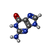
Mass: 151.126 Da / Num. of mol.: 4 / Source method: obtained synthetically / Formula: C5H5N5O / Feature type: SUBJECT OF INVESTIGATION
#3: Chemical
ChemComp-EMC / ETHYL MERCURY ION


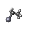
Mass: 229.651 Da / Num. of mol.: 4 / Source method: obtained synthetically / Formula: C2H5Hg
#4: Chemical ChemComp-PO4 / PHOSPHATE ION


Mass: 94.971 Da / Num. of mol.: 2 / Source method: obtained synthetically / Formula: PO4
#5: Chemical ChemComp-CA / CALCIUM ION


Mass: 40.078 Da / Num. of mol.: 1 / Source method: isolated from a natural source / Formula: Ca
#6: Water ChemComp-HOH / water


Mass: 18.015 Da / Num. of mol.: 648 / Source method: isolated from a natural source / Formula: H2O

-
Details

Has ligand of interestY

-
Experimental details

-
Experiment

ExperimentMethod: X-RAY DIFFRACTION / Number of used crystals: 1

-
Sample preparation

CrystalDensity Matthews: 2.58 Å3/Da / Density % sol: 52.24 %
Crystal growTemperature: 293 K / Method: vapor diffusion, sitting drop
Details: Protein at 20 mg/mL in SD2 plates incubated at 20 C. Conditions contained 6-8% Jeffamine M600 with 2-7% MPD and trisodium citrate at 1.1-1.2 M

-
Data collection

DiffractionMean temperature: 100 K / Serial crystal experiment: N
Diffraction sourceSource: SYNCHROTRON / Site: Australian Synchrotron / Beamline: MX2 / Wavelength: 1.00802 Å
DetectorType: DECTRIS EIGER X 16M / Detector: PIXEL / Date: Sep 26, 2018
RadiationProtocol: SINGLE WAVELENGTH / Monochromatic (M) / Laue (L): M / Scattering type: x-ray
Radiation wavelengthWavelength: 1.00802 Å / Relative weight: 1
ReflectionResolution: 1.87→49.4 Å / Num. obs: 119947 / % possible obs: 97.8 % / Redundancy: 19.6 % / CC1/2: 0.998 / Rmerge(I) obs: 0.14 / Rpim(I) all: 0.032 / Net I/σ(I): 13.5
Reflection shellResolution: 1.87→1.9 Å / Redundancy: 7.4 % / Rmerge(I) obs: 0.669 / Mean I/σ(I) obs: 3.1 / Num. unique obs: 4833 / CC1/2: 0.809 / Rpim(I) all: 0.246 / % possible all: 80.1

-
Processing

Software
NameVersionClassification
REFMAC5.8.0238refinement
ADDREFdata reduction
Aimlessdata scaling
CRANK2phasing
RefinementMethod to determine structure: SAD / Resolution: 1.87→49.4 Å / Cor.coef. Fo:Fc: 0.972 / Cor.coef. Fo:Fc free: 0.961 / SU B: 2.469 / SU ML: 0.073 / Cross valid method: THROUGHOUT / ESU R: 0.116 / ESU R Free: 0.108 / Details: HYDROGENS HAVE BEEN ADDED IN THE RIDING POSITIONS
RfactorNum. reflection% reflectionSelection details
Rfree0.18303 5791 4.8 %RANDOM
Rwork0.15486 ---
obs0.15624 114140 97.59 %-
Solvent computationIon probe radii: 0.8 Å / Shrinkage radii: 0.8 Å / VDW probe radii: 1.2 Å
Displacement parametersBiso mean: 32.759 Å2
Baniso -1Baniso -2Baniso -3
1-1.68 Å2-0 Å22.47 Å2
2---2 Å2-0 Å2
3---0.02 Å2
Refinement stepCycle: 1 / Resolution: 1.87→49.4 Å
ProteinNucleic acidLigandSolventTotal
Num. atoms10065 0 67 648 10780
Refine LS restraints
Refine-IDTypeDev idealDev ideal targetNumber
X-RAY DIFFRACTIONr_bond_refined_d0.0110.01310464
X-RAY DIFFRACTIONr_bond_other_d0.0010.0179771
X-RAY DIFFRACTIONr_angle_refined_deg1.5481.64114202
X-RAY DIFFRACTIONr_angle_other_deg1.4121.57922724
X-RAY DIFFRACTIONr_dihedral_angle_1_deg5.82251348
X-RAY DIFFRACTIONr_dihedral_angle_2_deg33.01824.009434
X-RAY DIFFRACTIONr_dihedral_angle_3_deg14.805151722
X-RAY DIFFRACTIONr_dihedral_angle_4_deg11.2291529
X-RAY DIFFRACTIONr_chiral_restr0.0770.21349
X-RAY DIFFRACTIONr_gen_planes_refined0.0090.0211784
X-RAY DIFFRACTIONr_gen_planes_other0.0010.022071
X-RAY DIFFRACTIONr_nbd_refined
X-RAY DIFFRACTIONr_nbd_other
X-RAY DIFFRACTIONr_nbtor_refined
X-RAY DIFFRACTIONr_nbtor_other
X-RAY DIFFRACTIONr_xyhbond_nbd_refined
X-RAY DIFFRACTIONr_xyhbond_nbd_other
X-RAY DIFFRACTIONr_metal_ion_refined
X-RAY DIFFRACTIONr_metal_ion_other
X-RAY DIFFRACTIONr_symmetry_vdw_refined
X-RAY DIFFRACTIONr_symmetry_vdw_other
X-RAY DIFFRACTIONr_symmetry_hbond_refined
X-RAY DIFFRACTIONr_symmetry_hbond_other
X-RAY DIFFRACTIONr_symmetry_metal_ion_refined
X-RAY DIFFRACTIONr_symmetry_metal_ion_other
X-RAY DIFFRACTIONr_mcbond_it2.8723.1835374
X-RAY DIFFRACTIONr_mcbond_other2.8713.1835373
X-RAY DIFFRACTIONr_mcangle_it3.6984.7546728
X-RAY DIFFRACTIONr_mcangle_other3.6974.7556729
X-RAY DIFFRACTIONr_scbond_it4.4353.6375090
X-RAY DIFFRACTIONr_scbond_other4.4053.6345082
X-RAY DIFFRACTIONr_scangle_it
X-RAY DIFFRACTIONr_scangle_other6.5995.2177463
X-RAY DIFFRACTIONr_long_range_B_refined7.08961.14845465
X-RAY DIFFRACTIONr_long_range_B_other7.07661.06445053
X-RAY DIFFRACTIONr_rigid_bond_restr
X-RAY DIFFRACTIONr_sphericity_free
X-RAY DIFFRACTIONr_sphericity_bonded
Refine LS restraints NCS

Refine-ID: X-RAY DIFFRACTION / Type: interatomic distance / Weight position: 0.05

Ens-IDDom-IDAuth asym-IDNumberRms dev position (Å)
11A108350.05
12B108350.05
21A105650.08
22C105650.08
31A105750.07
32D105750.07
41B105850.08
42C105850.08
51B105950.08
52D105950.08
61C106360.06
62D106360.06
LS refinement shellResolution: 1.869→1.918 Å / Total num. of bins used: 20
RfactorNum. reflection% reflection
Rfree0.274 374 -
Rwork0.228 7356 -
obs--85.65 %

+
About Yorodumi

-
News

-
Feb 9, 2022. New format data for meta-information of EMDB entries

New format data for meta-information of EMDB entries

  • Version 3 of the EMDB header file is now the official format.
  • The previous official version 1.9 will be removed from the archive.

Related info.:EMDB header

External links:wwPDB to switch to version 3 of the EMDB data model

-
Aug 12, 2020. Covid-19 info

Covid-19 info

URL: https://pdbjlvh1.pdbj.org/emnavi/covid19.php

New page: Covid-19 featured information page in EM Navigator.

Related info.:Covid-19 info / Mar 5, 2020. Novel coronavirus structure data

+
Mar 5, 2020. Novel coronavirus structure data

Novel coronavirus structure data

Related info.:Yorodumi Speices / Aug 12, 2020. Covid-19 info

External links:COVID-19 featured content - PDBj / Molecule of the Month (242):Coronavirus Proteases

+
Jan 31, 2019. EMDB accession codes are about to change! (news from PDBe EMDB page)

EMDB accession codes are about to change! (news from PDBe EMDB page)

  • The allocation of 4 digits for EMDB accession codes will soon come to an end. Whilst these codes will remain in use, new EMDB accession codes will include an additional digit and will expand incrementally as the available range of codes is exhausted. The current 4-digit format prefixed with “EMD-” (i.e. EMD-XXXX) will advance to a 5-digit format (i.e. EMD-XXXXX), and so on. It is currently estimated that the 4-digit codes will be depleted around Spring 2019, at which point the 5-digit format will come into force.
  • The EM Navigator/Yorodumi systems omit the EMD- prefix.

Related info.:Q: What is EMD? / ID/Accession-code notation in Yorodumi/EM Navigator

External links:EMDB Accession Codes are Changing Soon! / Contact to PDBj

+
Jul 12, 2017. Major update of PDB

Major update of PDB

  • wwPDB released updated PDB data conforming to the new PDBx/mmCIF dictionary.
  • This is a major update changing the version number from 4 to 5, and with Remediation, in which all the entries are updated.
  • In this update, many items about electron microscopy experimental information are reorganized (e.g. em_software).
  • Now, EM Navigator and Yorodumi are based on the updated data.

External links:wwPDB Remediation / Enriched Model Files Conforming to OneDep Data Standards Now Available in the PDB FTP Archive

-
Yorodumi

Thousand views of thousand structures

  • Yorodumi is a browser for structure data from EMDB, PDB, SASBDB, etc.
  • This page is also the successor to EM Navigator detail page, and also detail information page/front-end page for Omokage search.
  • The word "yorodu" (or yorozu) is an old Japanese word meaning "ten thousand". "mi" (miru) is to see.

Related info.:EMDB / PDB / SASBDB / Comparison of 3 databanks / Yorodumi Search / Aug 31, 2016. New EM Navigator & Yorodumi / Yorodumi Papers / Jmol/JSmol / Function and homology information / Changes in new EM Navigator and Yorodumi

Read more