 Movie
Movie Controller

[English] 日本語
 Yorodumi
- PDB-1o70: Novel Fold Revealed by the Structure of a FAS1 Domain Pair from t... -
+ Open data

- Basic information
| Entry | Database: PDB / ID: 1o70 | |||||||||
|---|---|---|---|---|---|---|---|---|---|---|
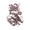
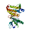
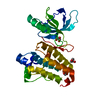
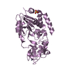
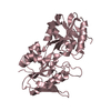
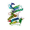
| Title | Novel Fold Revealed by the Structure of a FAS1 Domain Pair from the Insect Cell Adhesion Molecule Fasciclin I | |||||||||
 Components Components | FASCICLIN I | |||||||||
 Keywords Keywords | CELL ADHESION / AXON GUIDANCE / EXTRACELLULAR MODULE / GENETIC DISORDER / CORNEAL DYSTROPHY | |||||||||
| Function / homology |  Function and homology information Function and homology informationneuron recognition / calcium-independent cell-cell adhesion / homophilic cell-cell adhesion / side of membrane / extracellular matrix organization / cell adhesion molecule binding / axon guidance / extracellular matrix / cell adhesion / : / plasma membrane Similarity search - Function | |||||||||
| Biological species |  | |||||||||
| Method |  X-RAY DIFFRACTION / SYNCHROTRON / MIR / Resolution: 2.6 Å X-RAY DIFFRACTION / SYNCHROTRON / MIR / Resolution: 2.6 Å | |||||||||
 Authors Authors | Clout, N.J. / Tisi, D. / Hohenester, E. | |||||||||
 Citation Citation | Journal: Structure / Year: 2003 Title: Novel Fold Revealed by the Structure of a Fas1 Domain Pair from the Insect Cell Adhesion Molecule Fasciclin I Authors: Clout, N.J. / Tisi, D. / Hohenester, E. | |||||||||
| History |
|
- Structure visualization
| Structure viewer | Molecule: MolmilJmol/JSmol |
|---|
- Downloads & links
-Download
| PDBx/mmCIF format | 1o70.cif.gz | 75.8 KB | Display | PDBx/mmCIF format |
|---|---|---|---|---|
| PDB format | pdb1o70.ent.gz | 56.4 KB | Display | PDB format |
| PDBx/mmJSON format | 1o70.json.gz | Tree view | PDBx/mmJSON format | |
| Others |  Other downloads Other downloads |
-Validation report
| Arichive directory | https://data.pdbj.org/pub/pdb/validation_reports/o7/1o70ftp://data.pdbj.org/pub/pdb/validation_reports/o7/1o70 | HTTPS FTP |
|---|
-Related structure data
| Similar structure data |
|---|


-Links
 PDBj
PDBj- Assembly
| Deposited unit | 
| ||||||||
|---|---|---|---|---|---|---|---|---|---|
| 1 |
| ||||||||
| Unit cell |
|
-Components
| #1: Protein | Mass: 36493.141 Da / Num. of mol.: 1 / Fragment: FAS I DOMAINS 3 AND 4, RESIDUES 314-628 Source method: isolated from a genetically manipulated source Source: (gene. exp.)  HOMO SAPIENS (human) / References: UniProt: P10674 HOMO SAPIENS (human) / References: UniProt: P10674 | ||||||
|---|---|---|---|---|---|---|---|
| #2: Polysaccharide | 2-acetamido-2-deoxy-beta-D-glucopyranose-(1-4)-2-acetamido-2-deoxy-beta-D-glucopyranose Source method: isolated from a genetically manipulated source | ||||||
| #3: Chemical | ChemComp-SO4 /   Mass: 96.063 Da / Num. of mol.: 4 / Source method: obtained synthetically / Formula: SO4 Mass: 96.063 Da / Num. of mol.: 4 / Source method: obtained synthetically / Formula: SO4#4: Water | ChemComp-HOH / |  Mass: 18.015 Da / Num. of mol.: 76 / Source method: isolated from a natural source / Formula: H2O Mass: 18.015 Da / Num. of mol.: 76 / Source method: isolated from a natural source / Formula: H2OCompound details | NEURAL CELL ADHESION MOLECULE THAT IS EXPRESSED ON DIFFERENT SUBSETS OF AXON BUNDLES IN INSECT EMBRYOS. | Has protein modification | Y | |
-Experimental details
-Experiment
| Experiment | Method: X-RAY DIFFRACTION / Number of used crystals: 1 |
|---|
- Sample preparation
| Crystal | Density Matthews: 4.07 Å3/Da / Density % sol: 68 % | ||||||||||||||||||||||||||||||
|---|---|---|---|---|---|---|---|---|---|---|---|---|---|---|---|---|---|---|---|---|---|---|---|---|---|---|---|---|---|---|---|
| Crystal grow | pH: 8.5 / Details: 0.1 M TRIS-HCL PH 8.5, 2.0 M AMMONIUM SULFATE | ||||||||||||||||||||||||||||||
| Crystal grow | *PLUS pH: 7.5 / Method: vapor diffusion, hanging drop | ||||||||||||||||||||||||||||||
| Components of the solutions | *PLUS
|
-Data collection
| Diffraction | Mean temperature: 100 K |
|---|---|
| Diffraction source | Source: SYNCHROTRON / Site: SRS  / Beamline: PX9.6 / Wavelength: 0.87 / Beamline: PX9.6 / Wavelength: 0.87 |
| Detector | Type: ADSC CCD / Detector: CCD / Date: May 15, 2002 / Details: RH COATED SI MIRROR |
| Radiation | Monochromator: A TRIANGULAR SINGLE CRYSTAL SI MONOCHROMATOR / Protocol: SINGLE WAVELENGTH / Monochromatic (M) / Laue (L): M / Scattering type: x-ray |
| Radiation wavelength | Wavelength: 0.87 Å / Relative weight: 1 |
| Reflection | Resolution: 2.6→20 Å / Num. obs: 17949 / % possible obs: 97.1 % / Redundancy: 8.4 % / Rmerge(I) obs: 0.058 / Net I/σ(I): 4.4 |
| Reflection shell | Resolution: 2.6→20 Å / Redundancy: 8.3 % / Rmerge(I) obs: 0.31 / Mean I/σ(I) obs: 2.4 / % possible all: 97.5 |
| Reflection | *PLUS Lowest resolution: 20 Å |
| Reflection shell | *PLUS Highest resolution: 2.6 Å / Lowest resolution: 2.74 Å / % possible obs: 97.5 % / Rmerge(I) obs: 0.309 |
- Processing
| Software |
| ||||||||||||||||||||||||||||||||||||||||||||||||||||||||||||
|---|---|---|---|---|---|---|---|---|---|---|---|---|---|---|---|---|---|---|---|---|---|---|---|---|---|---|---|---|---|---|---|---|---|---|---|---|---|---|---|---|---|---|---|---|---|---|---|---|---|---|---|---|---|---|---|---|---|---|---|---|---|
| Refinement | Method to determine structure: MIR / Resolution: 2.6→20 Å / Data cutoff high absF: 10000 / Isotropic thermal model: RESTRAINED / Cross valid method: THROUGHOUT
| ||||||||||||||||||||||||||||||||||||||||||||||||||||||||||||
| Displacement parameters | Biso mean: 32.2 Å2 | ||||||||||||||||||||||||||||||||||||||||||||||||||||||||||||
| Refinement step | Cycle: LAST / Resolution: 2.6→20 Å
| ||||||||||||||||||||||||||||||||||||||||||||||||||||||||||||
| Refine LS restraints |
| ||||||||||||||||||||||||||||||||||||||||||||||||||||||||||||
| Xplor file |
| ||||||||||||||||||||||||||||||||||||||||||||||||||||||||||||
| Refinement | *PLUS Highest resolution: 2.6 Å / Lowest resolution: 20 Å / Num. reflection Rfree: 1758 / Rfactor Rfree: 0.263 | ||||||||||||||||||||||||||||||||||||||||||||||||||||||||||||
| Solvent computation | *PLUS | ||||||||||||||||||||||||||||||||||||||||||||||||||||||||||||
| Displacement parameters | *PLUS | ||||||||||||||||||||||||||||||||||||||||||||||||||||||||||||
| Refine LS restraints | *PLUS
|
