 Movie
Movie Controller

[English] 日本語
 Yorodumi
- PDB-1szd: Structural basis for nicotinamide cleavage and ADP-ribose transfe... -
+ Open data

- Basic information
| Entry | Database: PDB / ID: 1szd | ||||||
|---|---|---|---|---|---|---|---|
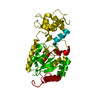
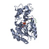
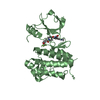
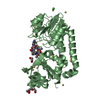
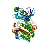
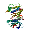
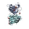
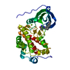
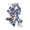
| Title | Structural basis for nicotinamide cleavage and ADP-ribose transfer by NAD+-dependent Sir2 histone/protein deacetylases | ||||||
 Components Components |
| ||||||
 Keywords Keywords | GENE REGULATION / hst2 / Sir2 / Sirtuin / histone deacetylase / protein deacetylase | ||||||
| Function / homology |  Function and homology information Function and homology information: / negative regulation of mitotic recombination / HATs acetylate histones / RNA polymerase I upstream activating factor complex / histone H4K16 deacetylase activity, NAD-dependent / Condensation of Prophase Chromosomes / : / : / : / Assembly of the ORC complex at the origin of replication ...: / negative regulation of mitotic recombination / HATs acetylate histones / RNA polymerase I upstream activating factor complex / histone H4K16 deacetylase activity, NAD-dependent / Condensation of Prophase Chromosomes / : / : / : / Assembly of the ORC complex at the origin of replication / HDACs deacetylate histones / protein acetyllysine N-acetyltransferase / histone deacetylase activity, NAD-dependent / rDNA heterochromatin formation / Recruitment and ATM-mediated phosphorylation of repair and signaling proteins at DNA double strand breaks / Oxidative Stress Induced Senescence / RMTs methylate histone arginines / SUMOylation of chromatin organization proteins / positive regulation of transcription by RNA polymerase I / RNA Polymerase I Promoter Escape / Estrogen-dependent gene expression / nucleolar large rRNA transcription by RNA polymerase I / NAD+ binding / structural constituent of chromatin / nucleosome / nucleosome assembly / chromatin organization / protein heterodimerization activity / regulation of DNA-templated transcription / DNA binding / metal ion binding / identical protein binding / nucleus / cytoplasm Similarity search - Function | ||||||
| Biological species |  | ||||||
| Method |  X-RAY DIFFRACTION / SYNCHROTRON / MOLECULAR REPLACEMENT / Resolution: 1.5 Å X-RAY DIFFRACTION / SYNCHROTRON / MOLECULAR REPLACEMENT / Resolution: 1.5 Å | ||||||
 Authors Authors | Zhao, K. / Harshaw, R. / Chai, X. / Marmorstein, R. | ||||||
 Citation Citation | Journal: Proc.Natl.Acad.Sci.Usa / Year: 2004 Title: Structural basis for nicotinamide cleavage and ADP-ribose transfer by NAD(+)-dependent Sir2 histone/protein deacetylases. Authors: Zhao, K. / Harshaw, R. / Chai, X. / Marmorstein, R. #1: Journal: Nat.Struct.Mol.Biol. / Year: 2003Title: Structure and Autoregulation Of The Yeast Hst2 Homolog Of Sir2 Authors: Zhao, K. / Harshaw, R. / Chai, X. / Marmorstein, R. #2: Journal: Structure / Year: 2003Title: Structure Of The Yeast Hst2 Protein Deacetylase In Ternary Complex With 2'-O-Acetyl ADP Ribose and Histone Peptide Authors: Zhao, K. / Chai, X. / Clements, A. / Marmorstein, R. | ||||||
| History |
|
- Structure visualization
| Structure viewer | Molecule: MolmilJmol/JSmol |
|---|
- Downloads & links
-Download
| PDBx/mmCIF format | 1szd.cif.gz | 86.1 KB | Display | PDBx/mmCIF format |
|---|---|---|---|---|
| PDB format | pdb1szd.ent.gz | 62.5 KB | Display | PDB format |
| PDBx/mmJSON format | 1szd.json.gz | Tree view | PDBx/mmJSON format | |
| Others |  Other downloads Other downloads |
-Validation report
| Arichive directory | https://data.pdbj.org/pub/pdb/validation_reports/sz/1szdftp://data.pdbj.org/pub/pdb/validation_reports/sz/1szd | HTTPS FTP |
|---|
-Related structure data
| Related structure data |  1szcC  1q1aS C: citing same article ( S: Starting model for refinement |
|---|---|
| Similar structure data |


-Links
 PDBj
PDBj


- Assembly
| Deposited unit | 
| ||||||||
|---|---|---|---|---|---|---|---|---|---|
| 1 |
| ||||||||
| 2 | 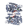
| ||||||||
| Unit cell |
|
-Components
-Protein / Protein/peptide , 2 types, 2 molecules AB
| #1: Protein | Mass: 33476.383 Da / Num. of mol.: 1 / Fragment: catalytic core domain Source method: isolated from a genetically manipulated source Source: (gene. exp.) Plasmid: hst2 (1-294) / Production host:  References: UniProt: P53686, Hydrolases; Acting on carbon-nitrogen bonds, other than peptide bonds; In linear amides |
|---|---|
| #2: Protein/peptide | Mass: 1197.457 Da / Num. of mol.: 1 / Source method: obtained synthetically Details: The peptide was chemically synthesized. The sequence of the peptide naturally occurs in Saccharomyces cerevisiae (baker's yeast). References: UniProt: P02309 |
-Non-polymers , 5 types, 346 molecules 


| #3: Chemical | ChemComp-CL /  Mass: 35.453 Da / Num. of mol.: 1 / Source method: obtained synthetically / Formula: Cl Mass: 35.453 Da / Num. of mol.: 1 / Source method: obtained synthetically / Formula: Cl | ||
|---|---|---|---|
| #4: Chemical | ChemComp-ZN /  Mass: 65.409 Da / Num. of mol.: 1 / Source method: obtained synthetically / Formula: Zn Mass: 65.409 Da / Num. of mol.: 1 / Source method: obtained synthetically / Formula: Zn | ||
| #5: Chemical | ChemComp-APR /  Mass: 559.316 Da / Num. of mol.: 1 / Source method: obtained synthetically / Formula: C15H23N5O14P2 Mass: 559.316 Da / Num. of mol.: 1 / Source method: obtained synthetically / Formula: C15H23N5O14P2 | ||
| #6: Chemical |  Mass: 92.094 Da / Num. of mol.: 3 / Source method: obtained synthetically / Formula: C3H8O3 Mass: 92.094 Da / Num. of mol.: 3 / Source method: obtained synthetically / Formula: C3H8O3#7: Water | ChemComp-HOH / | Mass: 18.015 Da / Num. of mol.: 340 / Source method: isolated from a natural source / Formula: H2O |
-Details
| Has protein modification | Y |
|---|
-Experimental details
-Experiment
| Experiment | Method: X-RAY DIFFRACTION / Number of used crystals: 1 |
|---|
- Sample preparation
| Crystal | Density Matthews: 3.2 Å3/Da / Density % sol: 61 % |
|---|---|
| Crystal grow | Temperature: 293 K / Method: vapor diffusion / pH: 6.5 Details: Ammonium Sulfate, Bis-Tris, pH 6.5, VAPOR DIFFUSION, temperature 293K |
-Data collection
| Diffraction | Mean temperature: 100 K |
|---|---|
| Diffraction source | Source: SYNCHROTRON / Site: NSLS  / Beamline: X25 / Wavelength: 0.9 Å / Beamline: X25 / Wavelength: 0.9 Å |
| Detector | Type: ADSC QUANTUM 4 / Detector: CCD / Date: Aug 8, 2003 |
| Radiation | Monochromator: Ni MIRROR + Ni FILTER / Protocol: SINGLE WAVELENGTH / Monochromatic (M) / Laue (L): M / Scattering type: x-ray |
| Radiation wavelength | Wavelength: 0.9 Å / Relative weight: 1 |
| Reflection | Resolution: 1.5→50 Å / Num. all: 67216 / Num. obs: 67216 / % possible obs: 99 % / Observed criterion σ(F): 2 / Observed criterion σ(I): 1 / Redundancy: 13 % / Biso Wilson estimate: 26.5 Å2 / Rmerge(I) obs: 0.063 / Rsym value: 0.059 / Net I/σ(I): 36.8 |
| Reflection shell | Resolution: 1.5→1.55 Å / Redundancy: 3 % / Rmerge(I) obs: 0.484 / Mean I/σ(I) obs: 3.3 / Num. unique all: 6325 / Rsym value: 0.437 / % possible all: 94.3 |
- Processing
| Software |
| ||||||||||||||||||||||||||||||||||||
|---|---|---|---|---|---|---|---|---|---|---|---|---|---|---|---|---|---|---|---|---|---|---|---|---|---|---|---|---|---|---|---|---|---|---|---|---|---|
| Refinement | Method to determine structure: MOLECULAR REPLACEMENT Starting model: PDB ENTRY 1Q1A Resolution: 1.5→28.01 Å / Rfactor Rfree error: 0.005 / Data cutoff high absF: 236729.23 / Data cutoff low absF: 0 / Isotropic thermal model: RESTRAINED / Cross valid method: THROUGHOUT / σ(F): 0 / Stereochemistry target values: Engh & Huber
| ||||||||||||||||||||||||||||||||||||
| Solvent computation | Solvent model: FLAT MODEL / Bsol: 60.6629 Å2 / ksol: 0.369871 e/Å3 | ||||||||||||||||||||||||||||||||||||
| Displacement parameters | Biso mean: 34.4 Å2
| ||||||||||||||||||||||||||||||||||||
| Refine analyze |
| ||||||||||||||||||||||||||||||||||||
| Refinement step | Cycle: LAST / Resolution: 1.5→28.01 Å
| ||||||||||||||||||||||||||||||||||||
| Refine LS restraints |
| ||||||||||||||||||||||||||||||||||||
| LS refinement shell | Resolution: 1.5→1.59 Å / Rfactor Rfree error: 0.02 / Total num. of bins used: 6
| ||||||||||||||||||||||||||||||||||||
| Xplor file |
|
