 Movie
Movie Controller


+ Open data

- Basic information
| Entry | Database: PDB / ID: 6pi6 | ||||||
|---|---|---|---|---|---|---|---|
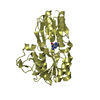
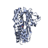
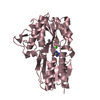
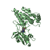
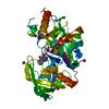
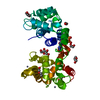
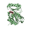
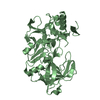
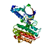
| Title | The evolving story of AtzT, a periplasmic binding protein | ||||||
 Components Components | Atrazine periplasmic binding protein | ||||||
 Keywords Keywords | TRANSPORT PROTEIN / SAD phasing / periplasmic binding protein / evolution from purine binding to atrazine binding | ||||||
| Function / homology |  Function and homology information Function and homology information | ||||||
| Biological species |  Pseudomonas sp. (bacteria) Pseudomonas sp. (bacteria) | ||||||
| Method |  X-RAY DIFFRACTION / SYNCHROTRON / MOLECULAR REPLACEMENT / Resolution: 1.65 Å X-RAY DIFFRACTION / SYNCHROTRON / MOLECULAR REPLACEMENT / Resolution: 1.65 Å | ||||||
 Authors Authors | Peat, T.S. / Newman, J. / Scott, C. / Esquirol, L. / Dennis, M. / Nebl, T. | ||||||
 Citation Citation | Journal: Acta Crystallogr D Struct Biol / Year: 2019 Title: The evolving story of AtzT, a periplasmic binding protein. Authors: Dennis, M.L. / Esquirol, L. / Nebl, T. / Newman, J. / Scott, C. / Peat, T.S. | ||||||
| History |
|
- Structure visualization
| Structure viewer | Molecule: MolmilJmol/JSmol |
|---|
- Downloads & links
-Download
| PDBx/mmCIF format | 6pi6.cif.gz | 281.2 KB | Display | PDBx/mmCIF format |
|---|---|---|---|---|
| PDB format | pdb6pi6.ent.gz | 224.5 KB | Display | PDB format |
| PDBx/mmJSON format | 6pi6.json.gz | Tree view | PDBx/mmJSON format | |
| Others |  Other downloads Other downloads |
-Validation report
| Arichive directory | https://data.pdbj.org/pub/pdb/validation_reports/pi/6pi6ftp://data.pdbj.org/pub/pdb/validation_reports/pi/6pi6 | HTTPS FTP |
|---|
-Related structure data
| Related structure data |  6pi5C  6piiSC S: Starting model for refinement C: citing same article ( |
|---|---|
| Similar structure data |

-Links
 PDBj
PDBj- Assembly
| Deposited unit | 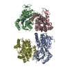
| ||||||||||||||||||||||||||||||||||||||||||||||||||||||||||||||||||||||||||||||||||||||||||||||||||||||||||||||||||||||||||||||||||||||||||||||||||||||
|---|---|---|---|---|---|---|---|---|---|---|---|---|---|---|---|---|---|---|---|---|---|---|---|---|---|---|---|---|---|---|---|---|---|---|---|---|---|---|---|---|---|---|---|---|---|---|---|---|---|---|---|---|---|---|---|---|---|---|---|---|---|---|---|---|---|---|---|---|---|---|---|---|---|---|---|---|---|---|---|---|---|---|---|---|---|---|---|---|---|---|---|---|---|---|---|---|---|---|---|---|---|---|---|---|---|---|---|---|---|---|---|---|---|---|---|---|---|---|---|---|---|---|---|---|---|---|---|---|---|---|---|---|---|---|---|---|---|---|---|---|---|---|---|---|---|---|---|---|---|---|---|
| 1 | 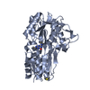
| ||||||||||||||||||||||||||||||||||||||||||||||||||||||||||||||||||||||||||||||||||||||||||||||||||||||||||||||||||||||||||||||||||||||||||||||||||||||
| 2 | 
| ||||||||||||||||||||||||||||||||||||||||||||||||||||||||||||||||||||||||||||||||||||||||||||||||||||||||||||||||||||||||||||||||||||||||||||||||||||||
| 3 | 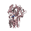
| ||||||||||||||||||||||||||||||||||||||||||||||||||||||||||||||||||||||||||||||||||||||||||||||||||||||||||||||||||||||||||||||||||||||||||||||||||||||
| 4 | 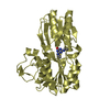
| ||||||||||||||||||||||||||||||||||||||||||||||||||||||||||||||||||||||||||||||||||||||||||||||||||||||||||||||||||||||||||||||||||||||||||||||||||||||
| Unit cell |
| ||||||||||||||||||||||||||||||||||||||||||||||||||||||||||||||||||||||||||||||||||||||||||||||||||||||||||||||||||||||||||||||||||||||||||||||||||||||
| Noncrystallographic symmetry (NCS) | NCS domain:
NCS domain segments: Component-ID: _ / Beg auth comp-ID: GLU / Beg label comp-ID: GLU / Refine code: _
NCS ensembles :
|
-Components
| #1: Protein | Mass: 38291.691 Da / Num. of mol.: 4 Source method: isolated from a genetically manipulated source Source: (gene. exp.) Pseudomonas sp. (strain ADP) (bacteria)Strain: ADP / Gene: orf97, AOX63_31690 / Production host: #2: Chemical | ChemComp-OKM /   Mass: 197.238 Da / Num. of mol.: 4 / Source method: obtained synthetically / Formula: C8H15N5O / Feature type: SUBJECT OF INVESTIGATION Mass: 197.238 Da / Num. of mol.: 4 / Source method: obtained synthetically / Formula: C8H15N5O / Feature type: SUBJECT OF INVESTIGATION#3: Chemical | ChemComp-DMS / |   Mass: 78.133 Da / Num. of mol.: 1 / Source method: obtained synthetically / Formula: C2H6OS / Comment: DMSO, precipitant*YM Mass: 78.133 Da / Num. of mol.: 1 / Source method: obtained synthetically / Formula: C2H6OS / Comment: DMSO, precipitant*YM#4: Water | ChemComp-HOH / |  Mass: 18.015 Da / Num. of mol.: 798 / Source method: isolated from a natural source / Formula: H2O Mass: 18.015 Da / Num. of mol.: 798 / Source method: isolated from a natural source / Formula: H2OHas ligand of interest | Y | |
|---|
-Experimental details
-Experiment
| Experiment | Method: X-RAY DIFFRACTION / Number of used crystals: 1 |
|---|
- Sample preparation
| Crystal | Density Matthews: 2.43 Å3/Da / Density % sol: 49.35 % |
|---|---|
| Crystal grow | Temperature: 293 K / Method: vapor diffusion, sitting drop Details: Protein at 20 mg/mL in SD2 plates incubated at 20 C. Crystallisation conditions contained 6-8% Jeffamine M600 with 2-7% MPD and trisodium citrate at 1.1-1.2 M |
-Data collection
| Diffraction | Mean temperature: 100 K / Serial crystal experiment: N |
|---|---|
| Diffraction source | Source: SYNCHROTRON / Site: Australian Synchrotron  / Beamline: MX2 / Wavelength: 0.953716 Å / Beamline: MX2 / Wavelength: 0.953716 Å |
| Detector | Type: DECTRIS EIGER X 16M / Detector: PIXEL / Date: Dec 18, 2018 |
| Radiation | Protocol: SINGLE WAVELENGTH / Monochromatic (M) / Laue (L): M / Scattering type: x-ray |
| Radiation wavelength | Wavelength: 0.953716 Å / Relative weight: 1 |
| Reflection | Resolution: 1.65→49.1 Å / Num. obs: 176707 / % possible obs: 100 % / Redundancy: 6.8 % / CC1/2: 0.998 / Rmerge(I) obs: 0.086 / Rpim(I) all: 0.035 / Net I/σ(I): 10.8 |
| Reflection shell | Resolution: 1.65→1.68 Å / Redundancy: 7 % / Rmerge(I) obs: 1.265 / Mean I/σ(I) obs: 1.5 / Num. unique obs: 8668 / CC1/2: 0.653 / Rpim(I) all: 0.515 / % possible all: 100 |
- Processing
| Software |
| |||||||||||||||||||||||||||||||||||||||||||||||||||||||||||||||||
|---|---|---|---|---|---|---|---|---|---|---|---|---|---|---|---|---|---|---|---|---|---|---|---|---|---|---|---|---|---|---|---|---|---|---|---|---|---|---|---|---|---|---|---|---|---|---|---|---|---|---|---|---|---|---|---|---|---|---|---|---|---|---|---|---|---|---|
| Refinement | Method to determine structure: MOLECULAR REPLACEMENT Starting model: 6PII Resolution: 1.65→49.1 Å / Cor.coef. Fo:Fc: 0.975 / Cor.coef. Fo:Fc free: 0.969 / SU B: 1.984 / SU ML: 0.063 / Cross valid method: THROUGHOUT / σ(F): 0 / ESU R: 0.082 / ESU R Free: 0.079 Details: HYDROGENS HAVE BEEN ADDED IN THE RIDING POSITIONS U VALUES : REFINED INDIVIDUALLY
| |||||||||||||||||||||||||||||||||||||||||||||||||||||||||||||||||
| Solvent computation | Ion probe radii: 0.8 Å / Shrinkage radii: 0.8 Å / VDW probe radii: 1.2 Å | |||||||||||||||||||||||||||||||||||||||||||||||||||||||||||||||||
| Displacement parameters | Biso max: 93.06 Å2 / Biso mean: 27.596 Å2 / Biso min: 14.75 Å2
| |||||||||||||||||||||||||||||||||||||||||||||||||||||||||||||||||
| Refinement step | Cycle: final / Resolution: 1.65→49.1 Å
| |||||||||||||||||||||||||||||||||||||||||||||||||||||||||||||||||
| Refine LS restraints |
| |||||||||||||||||||||||||||||||||||||||||||||||||||||||||||||||||
| Refine LS restraints NCS | Refine-ID: X-RAY DIFFRACTION / Type: interatomic distance / Weight position: 0.05
| |||||||||||||||||||||||||||||||||||||||||||||||||||||||||||||||||
| LS refinement shell | Resolution: 1.65→1.693 Å / Rfactor Rfree error: 0 / Total num. of bins used: 20
|
