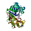
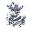
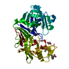
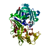
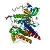
Entry Database : PDB / ID : 4itiTitle Crystal structure of RIP1 kinase in complex with necrostatin-3 analog Receptor-interacting serine/threonine-protein kinase 1 Keywords / / / / / / Function / homology Function Domain/homology Component
/ / / / / / / / / / / / / / / / / / / / / / / / / / / / / / / / / / / / / / / / / / / / / / / / / / / / / / / / / / / / / / / / / / / / / / / / / / / / / / / / / / / / / / / / / / / / / / / / / / / / / / / / / / / / / / / / / / / / / / / / / / / / / / / / / / / / / Biological species Homo sapiens (human)Method / / / Resolution : 2.86 Å Authors Xie, T. / Peng, W. / Liu, Y. / Yan, C. / Shi, Y. Journal : Structure / Year : 2013Title : Structural Basis of RIP1 Inhibition by Necrostatins.Authors : Xie, T. / Peng, W. / Liu, Y. / Yan, C. / Maki, J. / Degterev, A. / Yuan, J. / Shi, Y. History Deposition Jan 18, 2013 Deposition site / Processing site Revision 1.0 Mar 13, 2013 Provider / Type Revision 1.1 Mar 27, 2013 Group Revision 1.2 Sep 20, 2023 Group Data collection / Database references ... Data collection / Database references / Derived calculations / Refinement description Category chem_comp_atom / chem_comp_bond ... chem_comp_atom / chem_comp_bond / database_2 / pdbx_initial_refinement_model / struct_ref_seq_dif / struct_site Item _database_2.pdbx_DOI / _database_2.pdbx_database_accession ... _database_2.pdbx_DOI / _database_2.pdbx_database_accession / _struct_ref_seq_dif.details / _struct_site.pdbx_auth_asym_id / _struct_site.pdbx_auth_comp_id / _struct_site.pdbx_auth_seq_id
Show all Show less
 Movie
Movie Controller

 Yorodumi
 Open data
 Basic information
 Components
 Keywords
 Function and homology information
 Homo sapiens (human)
 X-RAY DIFFRACTION /
  Authors
 Citation
 Structure visualization
 Downloads & links
 Other downloads


 PDBj
PDBj


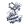
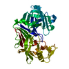
 Assembly


 Spodoptera frugiperda (fall armyworm)
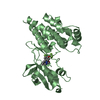
 Mass: 438.372 Da / Num. of mol.: 2 / Source method: obtained synthetically / Formula: C21H18F4N2O4
 Sample preparation
 / Beamline: BL17U / Wavelength: 0.9793 Å
 Processing