 Movie
Movie Controller

[English] 日本語
 Yorodumi
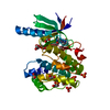
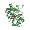
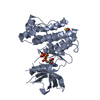
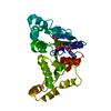
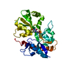
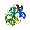
- PDB-4ith: Crystal structure of RIP1 kinase in complex with necrostatin-1 analog -
+ Open data

- Basic information
| Entry | Database: PDB / ID: 4ith | ||||||
|---|---|---|---|---|---|---|---|
| Title | Crystal structure of RIP1 kinase in complex with necrostatin-1 analog | ||||||
 Components Components | Receptor-interacting serine/threonine-protein kinase 1 | ||||||
 Keywords Keywords | TRANSFERASE/TRANSFERASE INHIBITOR / alpha/beta / RIP1 kinase / necroptosis / necrostatins / KINASE / TRANSFERASE-TRANSFERASE INHIBITOR complex | ||||||
| Function / homology |  Function and homology information Function and homology informationripoptosome assembly / positive regulation of miRNA processing / positive regulation of interleukin-6-mediated signaling pathway / death domain binding / ripoptosome assembly involved in necroptotic process / programmed necrotic cell death / Defective RIPK1-mediated regulated necrosis / Microbial modulation of RIPK1-mediated regulated necrosis / ripoptosome / TRIF-mediated programmed cell death ...ripoptosome assembly / positive regulation of miRNA processing / positive regulation of interleukin-6-mediated signaling pathway / death domain binding / ripoptosome assembly involved in necroptotic process / programmed necrotic cell death / Defective RIPK1-mediated regulated necrosis / Microbial modulation of RIPK1-mediated regulated necrosis / ripoptosome / TRIF-mediated programmed cell death / Regulation by c-FLIP / CASP8 activity is inhibited / Dimerization of procaspase-8 / TLR3-mediated TICAM1-dependent programmed cell death / Dengue virus modulates apoptosis / activation of protein kinase activity / positive regulation of macrophage differentiation / TNF signaling / Caspase activation via Death Receptors in the presence of ligand / T cell apoptotic process / SARS-CoV-1-mediated effects on programmed cell death / JUN kinase kinase kinase activity / necroptotic signaling pathway / NF-kB activation through FADD/RIP-1 pathway mediated by caspase-8 and -10 / negative regulation of necroptotic process / RIP-mediated NFkB activation via ZBP1 / death-inducing signaling complex / positive regulation of necroptotic process / positive regulation of tumor necrosis factor-mediated signaling pathway / death receptor binding / positive regulation of programmed cell death / positive regulation of programmed necrotic cell death / TNFR1-induced proapoptotic signaling / positive regulation of extrinsic apoptotic signaling pathway / RIPK1-mediated regulated necrosis / necroptotic process / TRP channels / extrinsic apoptotic signaling pathway via death domain receptors / protein serine/threonine phosphatase activity / response to tumor necrosis factor / positive regulation of execution phase of apoptosis / negative regulation of extrinsic apoptotic signaling pathway in absence of ligand / extrinsic apoptotic signaling pathway / canonical NF-kappaB signal transduction / signaling adaptor activity / negative regulation of extrinsic apoptotic signaling pathway / tumor necrosis factor-mediated signaling pathway / positive regulation of interleukin-8 production / negative regulation of canonical NF-kappaB signal transduction / TICAM1, RIP1-mediated IKK complex recruitment / protein serine/threonine kinase binding / IKK complex recruitment mediated by RIP1 / TNFR1-induced NF-kappa-B signaling pathway / Regulation of TNFR1 signaling / positive regulation of non-canonical NF-kappaB signal transduction / cellular response to growth factor stimulus / positive regulation of JNK cascade / intrinsic apoptotic signaling pathway in response to DNA damage / cellular response to tumor necrosis factor / Regulation of necroptotic cell death / positive regulation of reactive oxygen species metabolic process / cellular response to hydrogen peroxide / positive regulation of inflammatory response / positive regulation of tumor necrosis factor production / Ovarian tumor domain proteases / positive regulation of neuron apoptotic process / response to oxidative stress / amyloid fibril formation / Potential therapeutics for SARS / positive regulation of canonical NF-kappaB signal transduction / protein kinase activity / non-specific serine/threonine protein kinase / signaling receptor complex / endosome membrane / intracellular signal transduction / Ub-specific processing proteases / positive regulation of apoptotic process / inflammatory response / protein serine kinase activity / protein serine/threonine kinase activity / apoptotic process / positive regulation of gene expression / ubiquitin protein ligase binding / negative regulation of apoptotic process / protein-containing complex binding / protein homodimerization activity / positive regulation of transcription by RNA polymerase II / protein-containing complex / mitochondrion / ATP binding / identical protein binding / plasma membrane / cytosol / cytoplasm Similarity search - Function | ||||||
| Biological species |  Homo sapiens (human) Homo sapiens (human) | ||||||
| Method |  X-RAY DIFFRACTION / SYNCHROTRON / MOLECULAR REPLACEMENT / Resolution: 2.25 Å X-RAY DIFFRACTION / SYNCHROTRON / MOLECULAR REPLACEMENT / Resolution: 2.25 Å | ||||||
 Authors Authors | Xie, T. / Peng, W. / Liu, Y. / Yan, C. / Shi, Y. | ||||||
 Citation Citation | Journal: Structure / Year: 2013 Title: Structural Basis of RIP1 Inhibition by Necrostatins. Authors: Xie, T. / Peng, W. / Liu, Y. / Yan, C. / Maki, J. / Degterev, A. / Yuan, J. / Shi, Y. | ||||||
| History |
|
- Structure visualization
| Structure viewer | Molecule: MolmilJmol/JSmol |
|---|
- Downloads & links
-Download
| PDBx/mmCIF format | 4ith.cif.gz | 221.8 KB | Display | PDBx/mmCIF format |
|---|---|---|---|---|
| PDB format | pdb4ith.ent.gz | 178.6 KB | Display | PDB format |
| PDBx/mmJSON format | 4ith.json.gz | Tree view | PDBx/mmJSON format | |
| Others |  Other downloads Other downloads |
-Validation report
| Arichive directory | https://data.pdbj.org/pub/pdb/validation_reports/it/4ithftp://data.pdbj.org/pub/pdb/validation_reports/it/4ith | HTTPS FTP |
|---|
-Related structure data
| Related structure data |  4itiC  4itjC 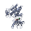 3c4cS C: citing same article ( S: Starting model for refinement |
|---|---|
| Similar structure data |


-Links
 PDBj
PDBj


- Assembly
| Deposited unit | 
| ||||||||
|---|---|---|---|---|---|---|---|---|---|
| 1 | 
| ||||||||
| 2 | 
| ||||||||
| Unit cell |
|
-Components
| #1: Protein | Mass: 33468.730 Da / Num. of mol.: 2 / Mutation: C34A, C127A, C233A, C240A Source method: isolated from a genetically manipulated source Source: (gene. exp.) Homo sapiens (human) / Gene: RIPK1, RIP, RIP1 / Production host:   Spodoptera frugiperda (fall armyworm) Spodoptera frugiperda (fall armyworm)References: UniProt: Q13546, non-specific serine/threonine protein kinase #2: Chemical | ChemComp-IOD /   Mass: 126.904 Da / Num. of mol.: 10 / Source method: obtained synthetically / Formula: I Mass: 126.904 Da / Num. of mol.: 10 / Source method: obtained synthetically / Formula: I#3: Chemical |   Mass: 22.990 Da / Num. of mol.: 2 / Source method: obtained synthetically / Formula: Na Mass: 22.990 Da / Num. of mol.: 2 / Source method: obtained synthetically / Formula: Na#4: Chemical |   Mass: 277.706 Da / Num. of mol.: 2 / Source method: obtained synthetically / Formula: C13H12ClN3O2 Mass: 277.706 Da / Num. of mol.: 2 / Source method: obtained synthetically / Formula: C13H12ClN3O2#5: Water | ChemComp-HOH / |  Mass: 18.015 Da / Num. of mol.: 132 / Source method: isolated from a natural source / Formula: H2O Mass: 18.015 Da / Num. of mol.: 132 / Source method: isolated from a natural source / Formula: H2O |
|---|
-Experimental details
-Experiment
| Experiment | Method: X-RAY DIFFRACTION / Number of used crystals: 1 |
|---|
- Sample preparation
| Crystal | Density Matthews: 2.13 Å3/Da / Density % sol: 42.16 % |
|---|---|
| Crystal grow | Temperature: 291 K / Method: vapor diffusion, hanging drop / pH: 5 Details: 0.25 M NH4I, 20% polyethylene glycol (PEG) 3350, 0.03 M glycyl-glycyl-glycine, pH 5.0, VAPOR DIFFUSION, HANGING DROP, temperature 291K |
-Data collection
| Diffraction | Mean temperature: 100 K |
|---|---|
| Diffraction source | Source: SYNCHROTRON / Site: SSRF  / Beamline: BL17U / Wavelength: 1 Å / Beamline: BL17U / Wavelength: 1 Å |
| Detector | Type: ADSC QUANTUM 315r / Detector: CCD / Date: Jan 11, 2012 |
| Radiation | Monochromator: Si 111 CHANNEL / Protocol: SINGLE WAVELENGTH / Monochromatic (M) / Laue (L): M / Scattering type: x-ray |
| Radiation wavelength | Wavelength: 1 Å / Relative weight: 1 |
| Reflection | Resolution: 2.25→40 Å / Num. all: 27731 / Num. obs: 27706 / % possible obs: 99.3 % / Observed criterion σ(F): 1 / Observed criterion σ(I): 1 |
| Reflection shell | Resolution: 2.25→2.33 Å / % possible all: 98.7 |
- Processing
| Software |
| |||||||||||||||||||||||||||||||||||||||||||||||||||||||||||||||||||||||||||||||||||||||||||||||||||||||||||||||||||||||||||||
|---|---|---|---|---|---|---|---|---|---|---|---|---|---|---|---|---|---|---|---|---|---|---|---|---|---|---|---|---|---|---|---|---|---|---|---|---|---|---|---|---|---|---|---|---|---|---|---|---|---|---|---|---|---|---|---|---|---|---|---|---|---|---|---|---|---|---|---|---|---|---|---|---|---|---|---|---|---|---|---|---|---|---|---|---|---|---|---|---|---|---|---|---|---|---|---|---|---|---|---|---|---|---|---|---|---|---|---|---|---|---|---|---|---|---|---|---|---|---|---|---|---|---|---|---|---|---|
| Refinement | Method to determine structure: MOLECULAR REPLACEMENT Starting model: PDB entry 3C4C Resolution: 2.25→37.888 Å / SU ML: 0.34 / σ(F): 0 / Phase error: 26.17 / Stereochemistry target values: ML
| |||||||||||||||||||||||||||||||||||||||||||||||||||||||||||||||||||||||||||||||||||||||||||||||||||||||||||||||||||||||||||||
| Solvent computation | Shrinkage radii: 1.11 Å / VDW probe radii: 1.3 Å / Solvent model: FLAT BULK SOLVENT MODEL / Bsol: 39.716 Å2 / ksol: 0.322 e/Å3 | |||||||||||||||||||||||||||||||||||||||||||||||||||||||||||||||||||||||||||||||||||||||||||||||||||||||||||||||||||||||||||||
| Displacement parameters |
| |||||||||||||||||||||||||||||||||||||||||||||||||||||||||||||||||||||||||||||||||||||||||||||||||||||||||||||||||||||||||||||
| Refinement step | Cycle: LAST / Resolution: 2.25→37.888 Å
| |||||||||||||||||||||||||||||||||||||||||||||||||||||||||||||||||||||||||||||||||||||||||||||||||||||||||||||||||||||||||||||
| Refine LS restraints |
| |||||||||||||||||||||||||||||||||||||||||||||||||||||||||||||||||||||||||||||||||||||||||||||||||||||||||||||||||||||||||||||
| LS refinement shell |
| |||||||||||||||||||||||||||||||||||||||||||||||||||||||||||||||||||||||||||||||||||||||||||||||||||||||||||||||||||||||||||||
| Refinement TLS params. | Method: refined / Refine-ID: X-RAY DIFFRACTION
| |||||||||||||||||||||||||||||||||||||||||||||||||||||||||||||||||||||||||||||||||||||||||||||||||||||||||||||||||||||||||||||
| Refinement TLS group |
|
