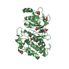
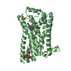
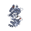
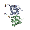
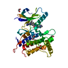
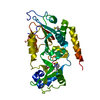
[English] 日本語
Yorodumi
- PDB-5f8u: Ligand occupancy in crystal structure of beta1-adrenergic recepto... -

+
Open data


ID or keywords:

Loading...

-
Basic information

Entry
Database: PDB / ID: 5f8u
TitleLigand occupancy in crystal structure of beta1-adrenergic receptor previously submitted by Huang et al
ComponentsBeta-1 adrenergic receptor
KeywordsSIGNALING PROTEIN / Beta1-AR / cyanopindolol
Function / homology
Function and homology information


beta1-adrenergic receptor activity / positive regulation of heart rate by epinephrine-norepinephrine / regulation of circadian sleep/wake cycle, sleep / norepinephrine-epinephrine-mediated vasodilation involved in regulation of systemic arterial blood pressure / adenylate cyclase-activating adrenergic receptor signaling pathway / early endosome / identical protein binding / membrane / plasma membrane
Similarity search - Function
Beta 1 adrenoceptor / Adrenoceptor family / Rhopdopsin 7-helix transmembrane proteins / Rhodopsin 7-helix transmembrane proteins / Serpentine type 7TM GPCR chemoreceptor Srsx / G-protein coupled receptors family 1 signature. / G protein-coupled receptor, rhodopsin-like / GPCR, rhodopsin-like, 7TM / G-protein coupled receptors family 1 profile. / 7 transmembrane receptor (rhodopsin family) ...Beta 1 adrenoceptor / Adrenoceptor family / Rhopdopsin 7-helix transmembrane proteins / Rhodopsin 7-helix transmembrane proteins / Serpentine type 7TM GPCR chemoreceptor Srsx / G-protein coupled receptors family 1 signature. / G protein-coupled receptor, rhodopsin-like / GPCR, rhodopsin-like, 7TM / G-protein coupled receptors family 1 profile. / 7 transmembrane receptor (rhodopsin family) / Up-down Bundle / Mainly Alpha
Similarity search - Domain/homology
Chem-P32 / Beta-1 adrenergic receptor
Similarity search - Component
Biological speciesMeleagris gallopavo (turkey)
MethodX-RAY DIFFRACTION / SYNCHROTRON / MOLECULAR REPLACEMENT / Resolution: 3.35 Å
AuthorsLeslie, A.G.W. / Warne, A. / Tate, C.G.
CitationJournal: Nat.Struct.Mol.Biol. / Year: 2013
Title: Crystal structure of oligomeric Beta1-adrenergic G protein-coupled receptors in ligand-free basal state.
Authors: Huang, J. / Chen, S. / Zhang, J.J. / Huang, X.Y.
History
DepositionDec 9, 2015Deposition site: RCSB / Processing site: PDBE
Revision 1.0Dec 23, 2015Provider: repository / Type: Initial release
Revision 1.1Mar 7, 2018Group: Data collection / Category: diffrn_source / Item: _diffrn_source.source
Revision 1.2Dec 26, 2018Group: Data collection / Experimental preparation / Category: exptl_crystal_grow / Item: _exptl_crystal_grow.method
Revision 1.3Apr 3, 2019Group: Data collection / Source and taxonomy / Category: entity_src_gen / Item: _entity_src_gen.pdbx_host_org_cell_line
Revision 1.4Jan 10, 2024Group: Data collection / Database references / Refinement description
Category: chem_comp_atom / chem_comp_bond ...chem_comp_atom / chem_comp_bond / database_2 / pdbx_initial_refinement_model / struct_ncs_dom_lim
Item: _database_2.pdbx_DOI / _database_2.pdbx_database_accession ..._database_2.pdbx_DOI / _database_2.pdbx_database_accession / _struct_ncs_dom_lim.beg_auth_comp_id / _struct_ncs_dom_lim.beg_label_asym_id / _struct_ncs_dom_lim.beg_label_comp_id / _struct_ncs_dom_lim.beg_label_seq_id / _struct_ncs_dom_lim.end_auth_comp_id / _struct_ncs_dom_lim.end_label_asym_id / _struct_ncs_dom_lim.end_label_comp_id / _struct_ncs_dom_lim.end_label_seq_id

-
Structure visualization

Structure viewerMolecule:
MolmilJmol/JSmol

Downloads & links

-
Assembly

Deposited unit
B: Beta-1 adrenergic receptor
A: Beta-1 adrenergic receptor
hetero molecules


Theoretical massNumber of molelcules
Total (without water)72,0804
Polymers71,5052
Non-polymers5752
Water00
1
B: Beta-1 adrenergic receptor
hetero molecules


Theoretical massNumber of molelcules
Total (without water)36,0402
Polymers35,7531
Non-polymers2871
Water0
TypeNameSymmetry operationNumber
identity operation1_555x,y,z1
2
A: Beta-1 adrenergic receptor
hetero molecules


Theoretical massNumber of molelcules
Total (without water)36,0402
Polymers35,7531
Non-polymers2871
Water0
TypeNameSymmetry operationNumber
identity operation1_555x,y,z1
Unit cell
Length a, b, c (Å)229.660, 79.590, 69.040
Angle α, β, γ (deg.)90.00, 101.83, 90.00
Int Tables number5
Space group name H-MC121
Noncrystallographic symmetry (NCS)NCS domain:
IDEns-IDDetails
11B
21A

NCS domain segments:

Component-ID: _ / Ens-ID: 1 / Beg auth comp-ID: GLN / Beg label comp-ID: GLN / End auth comp-ID: PHE / End label comp-ID: PHE / Refine code: _ / Auth seq-ID: 39 - 359 / Label seq-ID: 9 - 299

Dom-IDAuth asym-IDLabel asym-ID
1BA
2AB

-
Components

#1: Protein Beta-1 adrenergic receptor / Beta-1 adrenoreceptor / Beta-T


Mass: 35752.598 Da / Num. of mol.: 2 / Fragment: RESIDUES 33-243,272-276,279-367 / Mutation: R68S,M90V,Y227A,A282L,F327A,F338MC116L, C358A
Source method: isolated from a genetically manipulated source
Source: (gene. exp.) Meleagris gallopavo (turkey) / Gene: ADRB1 / Plasmid: PVL1393 REMA / Cell line (production host): High Five / Production host: 
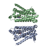
TRICHOPLUSIA NI (cabbage looper) / References: UniProt: P07700
#2: Chemical ChemComp-P32 / 4-{[(2S)-3-(tert-butylamino)-2-hydroxypropyl]oxy}-3H-indole-2-carbonitrile


Mass: 287.357 Da / Num. of mol.: 2 / Source method: obtained synthetically / Formula: C16H21N3O2

-
Experimental details

-
Experiment

ExperimentMethod: X-RAY DIFFRACTION / Number of used crystals: 1

-
Sample preparation

CrystalDensity Matthews: 4.95 Å3/Da / Density % sol: 75.13 % / Description: AUTHOR USED THE SF DATA FROM ENTRY 4GPO.

-
Data collection

DiffractionMean temperature: 100 K
Diffraction sourceSource: SYNCHROTRON / Site: NSLS / Beamline: X25 / Wavelength: 1 Å
DetectorType: PSI PILATUS 6M / Detector: PIXEL
RadiationProtocol: SINGLE WAVELENGTH / Monochromatic (M) / Laue (L): M / Scattering type: x-ray
Radiation wavelengthWavelength: 1 Å / Relative weight: 1
ReflectionResolution: 3.35→67.57 Å / Num. obs: 17463 / % possible obs: 98.2 % / Observed criterion σ(I): 2 / Redundancy: 4.3 % / Rmerge(I) obs: 0.141 / Net I/σ(I): 6.3
Reflection shellResolution: 3.35→3.44 Å / Redundancy: 4.2 % / Mean I/σ(I) obs: 1.7 / % possible all: 97

-
Processing

Software
NameVersionClassification
REFMAC5.8.0047refinement
XDSdata reduction
XSCALEdata scaling
RefinementMethod to determine structure: MOLECULAR REPLACEMENT
Starting model: 4gpo
Resolution: 3.35→67.57 Å / Cor.coef. Fo:Fc: 0.735 / Cor.coef. Fo:Fc free: 0.689 / SU B: 51.193 / SU ML: 0.79 / Cross valid method: THROUGHOUT / ESU R Free: 0.833 / Stereochemistry target values: MAXIMUM LIKELIHOOD / Details: HYDROGENS HAVE BEEN ADDED IN THE RIDING POSITIONS
RfactorNum. reflection% reflectionSelection details
Rfree0.38165 677 5 %RANDOM
Rwork0.34442 ---
obs0.34617 12909 76.51 %-
Solvent computationIon probe radii: 0.8 Å / Shrinkage radii: 0.8 Å / VDW probe radii: 1.2 Å / Solvent model: MASK
Displacement parametersBiso mean: 79.724 Å2
Baniso -1Baniso -2Baniso -3
1--0.19 Å20 Å2-0.28 Å2
2--1.79 Å2-0 Å2
3----1.36 Å2
Refinement stepCycle: LAST / Resolution: 3.35→67.57 Å
ProteinNucleic acidLigandSolventTotal
Num. atoms4390 0 42 0 4432
Refine LS restraints
Refine-IDTypeDev idealDev ideal targetNumber
X-RAY DIFFRACTIONr_bond_refined_d0.0130.024548
X-RAY DIFFRACTIONr_bond_other_d0.0040.024506
X-RAY DIFFRACTIONr_angle_refined_deg1.6291.9616210
X-RAY DIFFRACTIONr_angle_other_deg0.9983.00210256
X-RAY DIFFRACTIONr_dihedral_angle_1_deg6.2965548
X-RAY DIFFRACTIONr_dihedral_angle_2_deg37.37222.118170
X-RAY DIFFRACTIONr_dihedral_angle_3_deg22.80615742
X-RAY DIFFRACTIONr_dihedral_angle_4_deg11.2661530
X-RAY DIFFRACTIONr_chiral_restr0.10.2740
X-RAY DIFFRACTIONr_gen_planes_refined0.0080.024952
X-RAY DIFFRACTIONr_gen_planes_other0.0030.021090
X-RAY DIFFRACTIONr_nbd_refined
X-RAY DIFFRACTIONr_nbd_other
X-RAY DIFFRACTIONr_nbtor_refined
X-RAY DIFFRACTIONr_nbtor_other
X-RAY DIFFRACTIONr_xyhbond_nbd_refined
X-RAY DIFFRACTIONr_xyhbond_nbd_other
X-RAY DIFFRACTIONr_metal_ion_refined
X-RAY DIFFRACTIONr_metal_ion_other
X-RAY DIFFRACTIONr_symmetry_vdw_refined
X-RAY DIFFRACTIONr_symmetry_vdw_other
X-RAY DIFFRACTIONr_symmetry_hbond_refined
X-RAY DIFFRACTIONr_symmetry_hbond_other
X-RAY DIFFRACTIONr_symmetry_metal_ion_refined
X-RAY DIFFRACTIONr_symmetry_metal_ion_other
X-RAY DIFFRACTIONr_mcbond_it8.2327.5892204
X-RAY DIFFRACTIONr_mcbond_other8.2347.5822203
X-RAY DIFFRACTIONr_mcangle_it13.4111.352748
X-RAY DIFFRACTIONr_mcangle_other13.40811.3592749
X-RAY DIFFRACTIONr_scbond_it7.5348.4152337
X-RAY DIFFRACTIONr_scbond_other7.5328.4142338
X-RAY DIFFRACTIONr_scangle_it
X-RAY DIFFRACTIONr_scangle_other12.59512.3473457
X-RAY DIFFRACTIONr_long_range_B_refined25.20372.15720046
X-RAY DIFFRACTIONr_long_range_B_other25.20372.15520047
X-RAY DIFFRACTIONr_rigid_bond_restr
X-RAY DIFFRACTIONr_sphericity_free
X-RAY DIFFRACTIONr_sphericity_bonded
Refine LS restraints NCS

Ens-ID: 1 / Number: 17673 / Refine-ID: X-RAY DIFFRACTION / Type: interatomic distance / Rms dev position: 0.01 Å / Weight position: 0.05

Dom-IDAuth asym-ID
1B
2A
LS refinement shellResolution: 3.351→3.438 Å / Total num. of bins used: 20
RfactorNum. reflection% reflection
Rfree0.481 20 -
Rwork0.332 217 -
obs--18.08 %

+
About Yorodumi

-
News

-
Feb 9, 2022. New format data for meta-information of EMDB entries

New format data for meta-information of EMDB entries

  • Version 3 of the EMDB header file is now the official format.
  • The previous official version 1.9 will be removed from the archive.

Related info.:EMDB header

External links:wwPDB to switch to version 3 of the EMDB data model

-
Aug 12, 2020. Covid-19 info

Covid-19 info

URL: https://pdbjlvh1.pdbj.org/emnavi/covid19.php

New page: Covid-19 featured information page in EM Navigator.

Related info.:Covid-19 info / Mar 5, 2020. Novel coronavirus structure data

+
Mar 5, 2020. Novel coronavirus structure data

Novel coronavirus structure data

Related info.:Yorodumi Speices / Aug 12, 2020. Covid-19 info

External links:COVID-19 featured content - PDBj / Molecule of the Month (242):Coronavirus Proteases

+
Jan 31, 2019. EMDB accession codes are about to change! (news from PDBe EMDB page)

EMDB accession codes are about to change! (news from PDBe EMDB page)

  • The allocation of 4 digits for EMDB accession codes will soon come to an end. Whilst these codes will remain in use, new EMDB accession codes will include an additional digit and will expand incrementally as the available range of codes is exhausted. The current 4-digit format prefixed with “EMD-” (i.e. EMD-XXXX) will advance to a 5-digit format (i.e. EMD-XXXXX), and so on. It is currently estimated that the 4-digit codes will be depleted around Spring 2019, at which point the 5-digit format will come into force.
  • The EM Navigator/Yorodumi systems omit the EMD- prefix.

Related info.:Q: What is EMD? / ID/Accession-code notation in Yorodumi/EM Navigator

External links:EMDB Accession Codes are Changing Soon! / Contact to PDBj

+
Jul 12, 2017. Major update of PDB

Major update of PDB

  • wwPDB released updated PDB data conforming to the new PDBx/mmCIF dictionary.
  • This is a major update changing the version number from 4 to 5, and with Remediation, in which all the entries are updated.
  • In this update, many items about electron microscopy experimental information are reorganized (e.g. em_software).
  • Now, EM Navigator and Yorodumi are based on the updated data.

External links:wwPDB Remediation / Enriched Model Files Conforming to OneDep Data Standards Now Available in the PDB FTP Archive

-
Yorodumi

Thousand views of thousand structures

  • Yorodumi is a browser for structure data from EMDB, PDB, SASBDB, etc.
  • This page is also the successor to EM Navigator detail page, and also detail information page/front-end page for Omokage search.
  • The word "yorodu" (or yorozu) is an old Japanese word meaning "ten thousand". "mi" (miru) is to see.

Related info.:EMDB / PDB / SASBDB / Comparison of 3 databanks / Yorodumi Search / Aug 31, 2016. New EM Navigator & Yorodumi / Yorodumi Papers / Jmol/JSmol / Function and homology information / Changes in new EM Navigator and Yorodumi

Read more