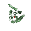
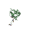
GAGPOLYPROTEIN, COREPROTEINP15 / EIAV MA / MATRIX PROTEIN / P15
Mass: 15022.591 Da / Num. of mol.: 2 / Fragment: MA PROTEIN Source method: isolated from a genetically manipulated source Details: THE PROTEIN HAS AN N-TERMINAL EXTENTION OF SEVEN RESIDUES DUE TO THE EXPRESSION CONSTRUCT. Source: (gene. exp.) EQUINE INFECTIOUS ANEMIA VIRUS / Strain: ISOLATE WYOMING / Gene: GAG / Variant: CLONE P3.2-1 / Plasmid: PET-32A / Production host: ESCHERICHIA COLI (E. coli) / Strain (production host): BL21(DE3) / References: UniProt: P03351, UniProt: P69732*PLUS
Has protein modification
Y
Sequence details
RESIDUES -6 TO 0 ARE A CLONING ARTIFACT
-
Experimental details
-
Experiment
Experiment
Method: X-RAY DIFFRACTION / Number of used crystals: 1
-
Sample preparation
Crystal
Density Matthews: 2.55 Å3/Da / Density % sol: 51.43 % / Description: FIGURES ARE SHOWN FOR THE WAVELENGTH 0.91116.
Crystal grow
Temperature: 291 K / pH: 6.5 Details: 30% PEG5000, 0.2M AMMONIUM SULFATE, 0.1M MES (PH6.5), 291K, pH 6.50
Crystal grow
*PLUS
Temperature: 18 ℃ / Method: vapor diffusion, sitting drop
Method to determine structure: MAD / Resolution: 2.8→23.78 Å / Rfactor Rfree error: 0.014 / Data cutoff low absF: 0 / Isotropic thermal model: RESTRAINED / Cross valid method: THROUGHOUT / σ(F): 0 Details: THE C-TERMINAL RESIDUES 110 TO 124 WERE NOT SEEN IN THE DENSITY MAPS
In the structure databanks used in Yorodumi, some data are registered as the other names, "COVID-19 virus" and "2019-nCoV". Here are the details of the virus and the list of structure data.
Jan 31, 2019. EMDB accession codes are about to change! (news from PDBe EMDB page)
EMDB accession codes are about to change! (news from PDBe EMDB page)
The allocation of 4 digits for EMDB accession codes will soon come to an end. Whilst these codes will remain in use, new EMDB accession codes will include an additional digit and will expand incrementally as the available range of codes is exhausted. The current 4-digit format prefixed with “EMD-” (i.e. EMD-XXXX) will advance to a 5-digit format (i.e. EMD-XXXXX), and so on. It is currently estimated that the 4-digit codes will be depleted around Spring 2019, at which point the 5-digit format will come into force.
The EM Navigator/Yorodumi systems omit the EMD- prefix.
Related info.:Q: What is EMD? / ID/Accession-code notation in Yorodumi/EM Navigator
Yorodumi is a browser for structure data from EMDB, PDB, SASBDB, etc.
This page is also the successor to EM Navigator detail page, and also detail information page/front-end page for Omokage search.
The word "yorodu" (or yorozu) is an old Japanese word meaning "ten thousand". "mi" (miru) is to see.
Related info.:EMDB / PDB / SASBDB / Comparison of 3 databanks / Yorodumi Search / Aug 31, 2016. New EM Navigator & Yorodumi / Yorodumi Papers / Jmol/JSmol / Function and homology information / Changes in new EM Navigator and Yorodumi
 Movie
Movie Controller

 Yorodumi
 Open data
 Basic information
 Components
 Keywords
 Function and homology information
 EQUINE INFECTIOUS ANEMIA VIRUS
 X-RAY DIFFRACTION /
  Authors
 Citation
 Structure visualization
 Downloads & links
 Other downloads


 PDBj
PDBj
 Assembly
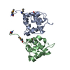


 Sample preparation
 / Beamline: BM14 / Wavelength: 0.9116, 0.9781, 0.9778
 Processing