positive regulation of meiotic cell cycle process involved in oocyte maturation / PKA activation in glucagon signalling / CREB1 phosphorylation through the activation of Adenylate Cyclase / DARPP-32 events / GPER1 signaling / Factors involved in megakaryocyte development and platelet production / PKA activation / Hedgehog 'off' state / regulation of meiotic cell cycle process involved in oocyte maturation / cAMP-dependent protein kinase regulator activity ...positive regulation of meiotic cell cycle process involved in oocyte maturation / PKA activation in glucagon signalling / CREB1 phosphorylation through the activation of Adenylate Cyclase / DARPP-32 events / GPER1 signaling / Factors involved in megakaryocyte development and platelet production / PKA activation / Hedgehog 'off' state / regulation of meiotic cell cycle process involved in oocyte maturation / cAMP-dependent protein kinase regulator activity / nucleotide-activated protein kinase complex / germinal vesicle / High laminar flow shear stress activates signaling by PIEZO1 and PECAM1:CDH5:KDR in endothelial cells / Vasopressin regulates renal water homeostasis via Aquaporins / cAMP-dependent protein kinase inhibitor activity / beta-2 adrenergic receptor binding / cAMP-dependent protein kinase complex / protein kinase A catalytic subunit binding / small molecule binding / protein kinase A binding / plasma membrane raft / axoneme / cAMP binding / negative regulation of cAMP/PKA signal transduction / T-tubule / modulation of chemical synaptic transmission / kinase activity / intracellular protein localization / adenylate cyclase-activating G protein-coupled receptor signaling pathway / protein domain specific binding / centrosome / ubiquitin protein ligase binding / synapse / protein-containing complex binding / perinuclear region of cytoplasm / glutamatergic synapse / protein-containing complex / mitochondrion / identical protein binding / plasma membrane / cytosol / cytoplasm Similarity search - Function
A-kinase anchor protein 10, PKA-binding (AKB) domain / : / cAMP-dependent protein kinase regulatory subunit, dimerization-anchoring domain / cAMP-dependent Protein Kinase, Chain A / cAMP-dependent protein kinase regulatory subunit / cAMP-dependent protein kinase regulatory subunit, dimerization-anchoring domain / Regulatory subunit of type II PKA R-subunit / RIIalpha, Regulatory subunit portion of type II PKA R-subunit / : / Regulator of G protein signaling domain ...A-kinase anchor protein 10, PKA-binding (AKB) domain / : / cAMP-dependent protein kinase regulatory subunit, dimerization-anchoring domain / cAMP-dependent Protein Kinase, Chain A / cAMP-dependent protein kinase regulatory subunit / cAMP-dependent protein kinase regulatory subunit, dimerization-anchoring domain / Regulatory subunit of type II PKA R-subunit / RIIalpha, Regulatory subunit portion of type II PKA R-subunit / : / Regulator of G protein signaling domain / RGS domain / RGS domain profile. / Regulator of G protein signalling domain / RGS, subdomain 2 / RGS domain superfamily / Cyclic nucleotide-binding domain signature 2. / Cyclic nucleotide-binding domain signature 1. / Cyclic nucleotide-binding, conserved site / Cyclic nucleotide-monophosphate binding domain / Cyclic nucleotide-binding domain / cAMP/cGMP binding motif profile. / Cyclic nucleotide-binding domain / Cyclic nucleotide-binding domain superfamily / RmlC-like jelly roll fold / Up-down Bundle / Mainly Alpha Similarity search - Domain/homology
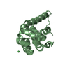
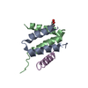
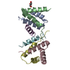
cAMP-dependent protein kinase type II-alpha regulatory subunit / A-kinase anchor protein 10, mitochondrial Similarity search - Component
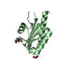
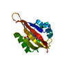
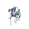
A: cAMP-dependent protein kinase type II-alpha regulatory subunit B: cAMP-dependent protein kinase type II-alpha regulatory subunit C: cAMP-dependent protein kinase type II-alpha regulatory subunit D: cAMP-dependent protein kinase type II-alpha regulatory subunit E: A Kinase binding peptide F: A Kinase binding peptide hetero molecules
A: cAMP-dependent protein kinase type II-alpha regulatory subunit B: cAMP-dependent protein kinase type II-alpha regulatory subunit E: A Kinase binding peptide hetero molecules
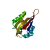
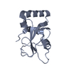
C: cAMP-dependent protein kinase type II-alpha regulatory subunit D: cAMP-dependent protein kinase type II-alpha regulatory subunit F: A Kinase binding peptide
A: cAMP-dependent protein kinase type II-alpha regulatory subunit B: cAMP-dependent protein kinase type II-alpha regulatory subunit E: A Kinase binding peptide hetero molecules
C: cAMP-dependent protein kinase type II-alpha regulatory subunit D: cAMP-dependent protein kinase type II-alpha regulatory subunit F: A Kinase binding peptide
In the structure databanks used in Yorodumi, some data are registered as the other names, "COVID-19 virus" and "2019-nCoV". Here are the details of the virus and the list of structure data.
Jan 31, 2019. EMDB accession codes are about to change! (news from PDBe EMDB page)
EMDB accession codes are about to change! (news from PDBe EMDB page)
The allocation of 4 digits for EMDB accession codes will soon come to an end. Whilst these codes will remain in use, new EMDB accession codes will include an additional digit and will expand incrementally as the available range of codes is exhausted. The current 4-digit format prefixed with “EMD-” (i.e. EMD-XXXX) will advance to a 5-digit format (i.e. EMD-XXXXX), and so on. It is currently estimated that the 4-digit codes will be depleted around Spring 2019, at which point the 5-digit format will come into force.
The EM Navigator/Yorodumi systems omit the EMD- prefix.
Related info.:Q: What is EMD? / ID/Accession-code notation in Yorodumi/EM Navigator
Yorodumi is a browser for structure data from EMDB, PDB, SASBDB, etc.
This page is also the successor to EM Navigator detail page, and also detail information page/front-end page for Omokage search.
The word "yorodu" (or yorozu) is an old Japanese word meaning "ten thousand". "mi" (miru) is to see.
Related info.:EMDB / PDB / SASBDB / Comparison of 3 databanks / Yorodumi Search / Aug 31, 2016. New EM Navigator & Yorodumi / Yorodumi Papers / Jmol/JSmol / Function and homology information / Changes in new EM Navigator and Yorodumi
 Movie
Movie Controller

 Yorodumi
 Open data
 Basic information
 Components
 Keywords
 Function and homology information
 X-RAY DIFFRACTION /
  Authors
 Citation
 Structure visualization
 Downloads & links
 Other downloads
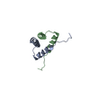


 PDBj
PDBj


 Assembly


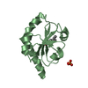
 Mass: 92.094 Da / Num. of mol.: 1 / Source method: obtained synthetically / Formula: C3H8O3
 Mass: 18.015 Da / Num. of mol.: 211 / Source method: isolated from a natural source / Formula: H2O
 Sample preparation
 / Beamline: 5.0.3 / Wavelength: 1 Å
 Processing