 Movie
Movie Controller


+ Open data

- Basic information
| Entry | Database: PDB / ID: 7aby | ||||||||||||
|---|---|---|---|---|---|---|---|---|---|---|---|---|---|
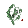
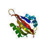
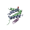
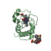
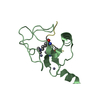
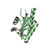
| Title | Crystal structure of iLOV-Q489K mutant | ||||||||||||
 Components Components | Phototropin-2 | ||||||||||||
 Keywords Keywords | SIGNALING PROTEIN / LOV domain / BLUE LIGHT PHOTORECEPTOR / FLAVOPROTEIN | ||||||||||||
| Function / homology |  Function and homology information Function and homology informationchloroplast relocation / phototropism / blue light signaling pathway / stomatal movement / blue light photoreceptor activity / response to blue light / plastid / circadian rhythm / kinase activity / FMN binding ...chloroplast relocation / phototropism / blue light signaling pathway / stomatal movement / blue light photoreceptor activity / response to blue light / plastid / circadian rhythm / kinase activity / FMN binding / non-specific serine/threonine protein kinase / protein serine kinase activity / protein serine/threonine kinase activity / Golgi apparatus / ATP binding / membrane / identical protein binding / nucleus / plasma membrane / cytoplasm Similarity search - Function | ||||||||||||
| Biological species |  | ||||||||||||
| Method |  X-RAY DIFFRACTION / SYNCHROTRON / MOLECULAR REPLACEMENT / molecular replacement / Resolution: 1.45 Å X-RAY DIFFRACTION / SYNCHROTRON / MOLECULAR REPLACEMENT / molecular replacement / Resolution: 1.45 Å | ||||||||||||
 Authors Authors | Granzin, J. / Batra-Safferling, R. | ||||||||||||
 Citation Citation | Journal: J.Biol.Chem. / Year: 2021 Title: The molecular basis of spectral tuning in blue- and red-shifted flavin-binding fluorescent proteins. Authors: Rollen, K. / Granzin, J. / Remeeva, A. / Davari, M.D. / Gensch, T. / Nazarenko, V.V. / Kovalev, K. / Bogorodskiy, A. / Borshchevskiy, V. / Hemmer, S. / Schwaneberg, U. / Gordeliy, V. / ...Authors: Rollen, K. / Granzin, J. / Remeeva, A. / Davari, M.D. / Gensch, T. / Nazarenko, V.V. / Kovalev, K. / Bogorodskiy, A. / Borshchevskiy, V. / Hemmer, S. / Schwaneberg, U. / Gordeliy, V. / Jaeger, K.E. / Batra-Safferling, R. / Gushchin, I. / Krauss, U. #1: Journal: Biorxiv / Year: 2021Title: Structural and mechanistic insight into spectral tuning in flavin-binding fluorescent proteins Authors: Rollen, K. / Granzin, J. / Remeeva, A. / Davari, M.D. / Gensch, T. / Nazarenko, V.V. / Kovalev, K. / Bogorodskiy, A. / Borshchevskiy, V. / Hemmer, S. / Schwaneberg, U. / Gordeliy, V. / ...Authors: Rollen, K. / Granzin, J. / Remeeva, A. / Davari, M.D. / Gensch, T. / Nazarenko, V.V. / Kovalev, K. / Bogorodskiy, A. / Borshchevskiy, V. / Hemmer, S. / Schwaneberg, U. / Gordeliy, V. / Jaeger, K.E. / Batra-Safferling, R. / Gushchin, I. / Krauss, U. | ||||||||||||
| History |
|
- Structure visualization
| Structure viewer | Molecule: MolmilJmol/JSmol |
|---|
- Downloads & links
-Download
| PDBx/mmCIF format | 7aby.cif.gz | 67.2 KB | Display | PDBx/mmCIF format |
|---|---|---|---|---|
| PDB format | pdb7aby.ent.gz | 46.2 KB | Display | PDB format |
| PDBx/mmJSON format | 7aby.json.gz | Tree view | PDBx/mmJSON format | |
| Others |  Other downloads Other downloads |
-Validation report
| Arichive directory | https://data.pdbj.org/pub/pdb/validation_reports/ab/7abyftp://data.pdbj.org/pub/pdb/validation_reports/ab/7aby | HTTPS FTP |
|---|
-Related structure data
| Related structure data |  6yx4C 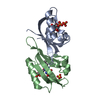 6yx6C 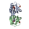 6yxbC 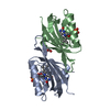 7ab6C 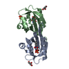 7ab7C 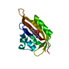 4eesS S: Starting model for refinement C: citing same article ( |
|---|---|
| Similar structure data |


-Links
 PDBj
PDBj


- Assembly
| Deposited unit | 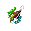
| ||||||||
|---|---|---|---|---|---|---|---|---|---|
| 1 |
| ||||||||
| Unit cell |
| ||||||||
| Components on special symmetry positions |
|
-Components
| #1: Protein | Mass: 15045.925 Da / Num. of mol.: 1 / Mutation: D491(SNN), G492(ACY), Q489K Source method: isolated from a genetically manipulated source Details: modified residues: Asp491-Gly492 to SNN L-3-AMINOSUCCINIMIDE and ACY ACETIC ACID including the required backbone bonds. Asp-Gly form a Succinimide intermediate. Keyword: Deamidation Source: (gene. exp.)  References: UniProt: P93025, non-specific serine/threonine protein kinase |
|---|---|
| #2: Chemical | ChemComp-FMN /   Mass: 456.344 Da / Num. of mol.: 1 / Source method: obtained synthetically / Formula: C17H21N4O9P / Feature type: SUBJECT OF INVESTIGATION Mass: 456.344 Da / Num. of mol.: 1 / Source method: obtained synthetically / Formula: C17H21N4O9P / Feature type: SUBJECT OF INVESTIGATION |
| #3: Chemical | ChemComp-ACT /   Mass: 59.044 Da / Num. of mol.: 1 / Source method: obtained synthetically / Formula: C2H3O2 Mass: 59.044 Da / Num. of mol.: 1 / Source method: obtained synthetically / Formula: C2H3O2 |
| #4: Water | ChemComp-HOH /  Mass: 18.015 Da / Num. of mol.: 106 / Source method: isolated from a natural source / Formula: H2O Mass: 18.015 Da / Num. of mol.: 106 / Source method: isolated from a natural source / Formula: H2O |
| Has ligand of interest | Y |
| Has protein modification | Y |
-Experimental details
-Experiment
| Experiment | Method: X-RAY DIFFRACTION / Number of used crystals: 1 |
|---|
- Sample preparation
| Crystal | Density Matthews: 1.82 Å3/Da / Density % sol: 32.56 % |
|---|---|
| Crystal grow | Temperature: 292.15 K / Method: vapor diffusion, sitting drop / pH: 4.6 / Details: 0.1 M sodium acetate, 25 % (w/v) PEG 1000 |
-Data collection
| Diffraction | Mean temperature: 100 K / Serial crystal experiment: N | ||||||||||||||||||||||||||||||
|---|---|---|---|---|---|---|---|---|---|---|---|---|---|---|---|---|---|---|---|---|---|---|---|---|---|---|---|---|---|---|---|
| Diffraction source | Source: SYNCHROTRON / Site: ESRF  / Beamline: ID29 / Wavelength: 0.9194 Å / Beamline: ID29 / Wavelength: 0.9194 Å | ||||||||||||||||||||||||||||||
| Detector | Type: DECTRIS PILATUS 6M-F / Detector: PIXEL / Date: Nov 27, 2017 | ||||||||||||||||||||||||||||||
| Radiation | Protocol: SINGLE WAVELENGTH / Monochromatic (M) / Laue (L): M / Scattering type: x-ray | ||||||||||||||||||||||||||||||
| Radiation wavelength | Wavelength: 0.9194 Å / Relative weight: 1 | ||||||||||||||||||||||||||||||
| Reflection | Resolution: 1.45→43.93 Å / Num. obs: 20710 / % possible obs: 100 % / Redundancy: 9 % / Biso Wilson estimate: 19.01 Å2 / CC1/2: 0.999 / Rmerge(I) obs: 0.07 / Rpim(I) all: 0.025 / Rrim(I) all: 0.074 / Net I/σ(I): 14.3 / Num. measured all: 186700 | ||||||||||||||||||||||||||||||
| Reflection shell | Diffraction-ID: 1
|
-Phasing
| Phasing | Method: molecular replacement | ||||||
|---|---|---|---|---|---|---|---|
| Phasing MR | R rigid body: 0.482
|
- Processing
| Software |
| ||||||||||||||||||||||||||||||||||||||||
|---|---|---|---|---|---|---|---|---|---|---|---|---|---|---|---|---|---|---|---|---|---|---|---|---|---|---|---|---|---|---|---|---|---|---|---|---|---|---|---|---|---|
| Refinement | Method to determine structure: MOLECULAR REPLACEMENT Starting model: 4EES Resolution: 1.45→38.937 Å / SU ML: 0.12 / Cross valid method: FREE R-VALUE / σ(F): 1.35 / Phase error: 17.14 / Stereochemistry target values: ML
| ||||||||||||||||||||||||||||||||||||||||
| Solvent computation | Shrinkage radii: 1 Å / VDW probe radii: 1.2 Å / Solvent model: FLAT BULK SOLVENT MODEL | ||||||||||||||||||||||||||||||||||||||||
| Displacement parameters | Biso max: 59.3 Å2 / Biso mean: 24.9037 Å2 / Biso min: 12.97 Å2 | ||||||||||||||||||||||||||||||||||||||||
| Refinement step | Cycle: final / Resolution: 1.45→38.937 Å
| ||||||||||||||||||||||||||||||||||||||||
| LS refinement shell | Refine-ID: X-RAY DIFFRACTION / Rfactor Rfree error: 0 / % reflection obs: 100 %
|
