 Movie
Movie Controller

[English] 日本語
 Yorodumi
- PDB-7ab7: Structure of Chloroflexus aggregans flavin based fluorescent prot... -
+ Open data

- Basic information
| Entry | Database: PDB / ID: 7ab7 | ||||||
|---|---|---|---|---|---|---|---|
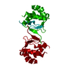
| Title | Structure of Chloroflexus aggregans flavin based fluorescent protein (CagFbFP) I52T Q148K variant | ||||||
 Components Components | Multi-sensor hybrid histidine kinase | ||||||
 Keywords Keywords | FLUORESCENT PROTEIN / LOV domain | ||||||
| Function / homology |  Function and homology information Function and homology informationphosphorelay sensor kinase activity / histidine kinase / ATP binding / plasma membrane Similarity search - Function | ||||||
| Biological species |   Chloroflexus aggregans (bacteria) Chloroflexus aggregans (bacteria) | ||||||
| Method |  X-RAY DIFFRACTION / SYNCHROTRON / MOLECULAR REPLACEMENT / Resolution: 1.8 Å X-RAY DIFFRACTION / SYNCHROTRON / MOLECULAR REPLACEMENT / Resolution: 1.8 Å | ||||||
 Authors Authors | Remeeva, A. / Nazarenko, V. / Kovalev, K. / Gushchin, I. | ||||||
 Citation Citation | Journal: J.Biol.Chem. / Year: 2021 Title: The molecular basis of spectral tuning in blue- and red-shifted flavin-binding fluorescent proteins. Authors: Rollen, K. / Granzin, J. / Remeeva, A. / Davari, M.D. / Gensch, T. / Nazarenko, V.V. / Kovalev, K. / Bogorodskiy, A. / Borshchevskiy, V. / Hemmer, S. / Schwaneberg, U. / Gordeliy, V. / ...Authors: Rollen, K. / Granzin, J. / Remeeva, A. / Davari, M.D. / Gensch, T. / Nazarenko, V.V. / Kovalev, K. / Bogorodskiy, A. / Borshchevskiy, V. / Hemmer, S. / Schwaneberg, U. / Gordeliy, V. / Jaeger, K.E. / Batra-Safferling, R. / Gushchin, I. / Krauss, U. #1: Journal: Biorxiv / Year: 2021Title: Structural and mechanistic insight into spectral tuning in flavin-binding fluorescent proteins Authors: Rollen, K. / Granzin, J. / Remeeva, A. / Davari, M.D. / Gensch, T. / Nazarenko, V.V. / Kovalev, K. / Bogorodskiy, A. / Borshchevskiy, V. / Hemmer, S. / Schwaneberg, U. / Gordeliy, V. / ...Authors: Rollen, K. / Granzin, J. / Remeeva, A. / Davari, M.D. / Gensch, T. / Nazarenko, V.V. / Kovalev, K. / Bogorodskiy, A. / Borshchevskiy, V. / Hemmer, S. / Schwaneberg, U. / Gordeliy, V. / Jaeger, K.E. / Batra-Safferling, R. / Gushchin, I. / Krauss, U. | ||||||
| History |
|
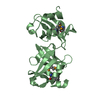
- Structure visualization
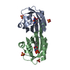
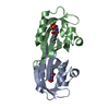
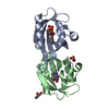
| Structure viewer | Molecule: MolmilJmol/JSmol |
|---|
- Downloads & links
-Download
| PDBx/mmCIF format | 7ab7.cif.gz | 64.9 KB | Display | PDBx/mmCIF format |
|---|---|---|---|---|
| PDB format | pdb7ab7.ent.gz | 46.2 KB | Display | PDB format |
| PDBx/mmJSON format | 7ab7.json.gz | Tree view | PDBx/mmJSON format | |
| Others |  Other downloads Other downloads |
-Validation report
| Arichive directory | https://data.pdbj.org/pub/pdb/validation_reports/ab/7ab7ftp://data.pdbj.org/pub/pdb/validation_reports/ab/7ab7 | HTTPS FTP |
|---|
-Related structure data
| Related structure data |  6yx4C 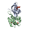 6yx6C 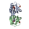 6yxbC 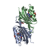 7ab6C 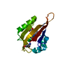 7abyC 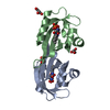 6rhfS S: Starting model for refinement C: citing same article ( |
|---|---|
| Similar structure data |


-Links
 PDBj
PDBj


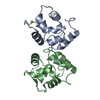
- Assembly
| Deposited unit | 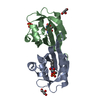
| ||||||||
|---|---|---|---|---|---|---|---|---|---|
| 1 |
| ||||||||
| Unit cell |
| ||||||||
| Components on special symmetry positions |
|
-Components
| #1: Protein | Mass: 12389.880 Da / Num. of mol.: 2 / Mutation: I52T Q148K Source method: isolated from a genetically manipulated source Source: (gene. exp.) Chloroflexus aggregans (strain MD-66 / DSM 9485) (bacteria)Strain: MD-66 / DSM 9485 / Gene: Cagg_3753 / Production host: #2: Chemical |   Mass: 456.344 Da / Num. of mol.: 2 / Source method: obtained synthetically / Formula: C17H21N4O9P / Feature type: SUBJECT OF INVESTIGATION Mass: 456.344 Da / Num. of mol.: 2 / Source method: obtained synthetically / Formula: C17H21N4O9P / Feature type: SUBJECT OF INVESTIGATION#3: Chemical |   Mass: 92.094 Da / Num. of mol.: 2 / Source method: obtained synthetically / Formula: C3H8O3 Mass: 92.094 Da / Num. of mol.: 2 / Source method: obtained synthetically / Formula: C3H8O3#4: Chemical | ChemComp-SO4 / |   Mass: 96.063 Da / Num. of mol.: 1 / Source method: obtained synthetically / Formula: SO4 Mass: 96.063 Da / Num. of mol.: 1 / Source method: obtained synthetically / Formula: SO4#5: Water | ChemComp-HOH / |  Mass: 18.015 Da / Num. of mol.: 175 / Source method: isolated from a natural source / Formula: H2O Mass: 18.015 Da / Num. of mol.: 175 / Source method: isolated from a natural source / Formula: H2OHas ligand of interest | Y | |
|---|
-Experimental details
-Experiment
| Experiment | Method: X-RAY DIFFRACTION / Number of used crystals: 1 |
|---|
- Sample preparation
| Crystal | Density Matthews: 2.56 Å3/Da / Density % sol: 51.89 % |
|---|---|
| Crystal grow | Temperature: 293 K / Method: vapor diffusion, sitting drop Details: 0.2M Lithium sulfate 0.1M BIS-Tris pH 5.5 25% PEG 3350 |
-Data collection
| Diffraction | Mean temperature: 100 K / Serial crystal experiment: N | ||||||||||||||||||||||||||||||
|---|---|---|---|---|---|---|---|---|---|---|---|---|---|---|---|---|---|---|---|---|---|---|---|---|---|---|---|---|---|---|---|
| Diffraction source | Source: SYNCHROTRON / Site: PETRA III, EMBL c/o DESY  / Beamline: P13 (MX1) / Wavelength: 0.9762 Å / Beamline: P13 (MX1) / Wavelength: 0.9762 Å | ||||||||||||||||||||||||||||||
| Detector | Type: DECTRIS PILATUS 6M / Detector: PIXEL / Date: May 20, 2019 | ||||||||||||||||||||||||||||||
| Radiation | Protocol: SINGLE WAVELENGTH / Monochromatic (M) / Laue (L): M / Scattering type: x-ray | ||||||||||||||||||||||||||||||
| Radiation wavelength | Wavelength: 0.9762 Å / Relative weight: 1 | ||||||||||||||||||||||||||||||
| Reflection | Resolution: 1.8→53.08 Å / Num. obs: 22158 / % possible obs: 99.5 % / Redundancy: 3.3 % / CC1/2: 0.996 / Rmerge(I) obs: 0.077 / Rpim(I) all: 0.049 / Rrim(I) all: 0.092 / Net I/σ(I): 7.1 / Num. measured all: 72891 / Scaling rejects: 1 | ||||||||||||||||||||||||||||||
| Reflection shell | Diffraction-ID: 1
|
- Processing
| Software |
| ||||||||||||||||||||||||||||||||||||||||||||||||||||||||||||
|---|---|---|---|---|---|---|---|---|---|---|---|---|---|---|---|---|---|---|---|---|---|---|---|---|---|---|---|---|---|---|---|---|---|---|---|---|---|---|---|---|---|---|---|---|---|---|---|---|---|---|---|---|---|---|---|---|---|---|---|---|---|
| Refinement | Method to determine structure: MOLECULAR REPLACEMENT Starting model: 6RHF Resolution: 1.8→48.01 Å / Cor.coef. Fo:Fc: 0.966 / Cor.coef. Fo:Fc free: 0.957 / SU B: 2.815 / SU ML: 0.085 / Cross valid method: THROUGHOUT / σ(F): 0 / ESU R: 0.114 / ESU R Free: 0.11 / Stereochemistry target values: MAXIMUM LIKELIHOOD Details: HYDROGENS HAVE BEEN ADDED IN THE RIDING POSITIONS U VALUES : REFINED INDIVIDUALLY
| ||||||||||||||||||||||||||||||||||||||||||||||||||||||||||||
| Solvent computation | Ion probe radii: 0.8 Å / Shrinkage radii: 0.8 Å / VDW probe radii: 1.2 Å / Solvent model: MASK | ||||||||||||||||||||||||||||||||||||||||||||||||||||||||||||
| Displacement parameters | Biso max: 68.67 Å2 / Biso mean: 21.224 Å2 / Biso min: 12.16 Å2
| ||||||||||||||||||||||||||||||||||||||||||||||||||||||||||||
| Refinement step | Cycle: final / Resolution: 1.8→48.01 Å
| ||||||||||||||||||||||||||||||||||||||||||||||||||||||||||||
| Refine LS restraints |
| ||||||||||||||||||||||||||||||||||||||||||||||||||||||||||||
| LS refinement shell | Resolution: 1.8→1.847 Å / Rfactor Rfree error: 0 / Total num. of bins used: 20
|
