 Movie
Movie Controller

[English] 日本語
 Yorodumi
- PDB-6ywg: Structure of Chloroflexus aggregans flavin based fluorescent prot... -
+ Open data

- Basic information
| Entry | Database: PDB / ID: 6ywg | ||||||
|---|---|---|---|---|---|---|---|
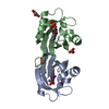
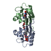
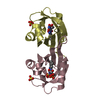
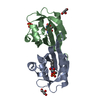
| Title | Structure of Chloroflexus aggregans flavin based fluorescent protein (CagFbFP) Q148N variant | ||||||
 Components Components | Multi-sensor hybrid histidine kinase | ||||||
 Keywords Keywords | FLUORESCENT PROTEIN / LOV domain | ||||||
| Function / homology |  Function and homology information Function and homology informationphosphorelay sensor kinase activity / histidine kinase / ATP binding / plasma membrane Similarity search - Function | ||||||
| Biological species |   Chloroflexus aggregans DSM 9485 (bacteria) Chloroflexus aggregans DSM 9485 (bacteria) | ||||||
| Method |  X-RAY DIFFRACTION / SYNCHROTRON / MOLECULAR REPLACEMENT / Resolution: 1.45 Å X-RAY DIFFRACTION / SYNCHROTRON / MOLECULAR REPLACEMENT / Resolution: 1.45 Å | ||||||
 Authors Authors | Remeeva, A. / Nazarenko, V. / Kovalev, K. / Gushchin, I. | ||||||
| Funding support |  Russian Federation, 1items Russian Federation, 1items
| ||||||
 Citation Citation | Journal: Proteins / Year: 2021 Title: Insights into the mechanisms of light-oxygen-voltage domain color tuning from a set of high-resolution X-ray structures. Authors: Remeeva, A. / Nazarenko, V.V. / Kovalev, K. / Goncharov, I.M. / Yudenko, A. / Astashkin, R. / Gordeliy, V. / Gushchin, I. #1: Journal: Biorxiv / Year: 2021Title: Insights into the mechanisms of LOV domain color tuning from a set of high-resolution X-ray structures Authors: Remeeva, A. / Nazarenko, V.V. / Kovalev, K. / Goncharov, I. / Yudenko, A. / Astashkin, R. / Gordeliy, V. / Gushchin, I. | ||||||
| History |
|
- Structure visualization
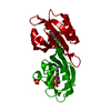
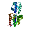
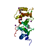
| Structure viewer | Molecule: MolmilJmol/JSmol |
|---|
- Downloads & links
-Download
| PDBx/mmCIF format | 6ywg.cif.gz | 65.2 KB | Display | PDBx/mmCIF format |
|---|---|---|---|---|
| PDB format | pdb6ywg.ent.gz | 46.9 KB | Display | PDB format |
| PDBx/mmJSON format | 6ywg.json.gz | Tree view | PDBx/mmJSON format | |
| Others |  Other downloads Other downloads |
-Validation report
| Arichive directory | https://data.pdbj.org/pub/pdb/validation_reports/yw/6ywgftp://data.pdbj.org/pub/pdb/validation_reports/yw/6ywg | HTTPS FTP |
|---|
-Related structure data
| Related structure data | 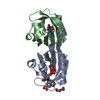 6ywhC 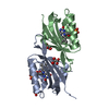 6ywiC 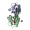 6ywqC 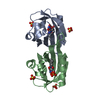 6ywrC 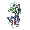 6yxcC 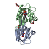 6rhfS S: Starting model for refinement C: citing same article ( |
|---|---|
| Similar structure data |


-Links
 PDBj
PDBj


- Assembly
| Deposited unit | 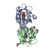
| ||||||||
|---|---|---|---|---|---|---|---|---|---|
| 1 |
| ||||||||
| Unit cell |
| ||||||||
| Components on special symmetry positions |
|
-Components
| #1: Protein | Mass: 12386.856 Da / Num. of mol.: 2 / Mutation: Q148N Source method: isolated from a genetically manipulated source Source: (gene. exp.) Chloroflexus aggregans DSM 9485 (bacteria)Gene: Cagg_3753 / Production host: #2: Chemical |   Mass: 456.344 Da / Num. of mol.: 2 / Source method: obtained synthetically / Formula: C17H21N4O9P Mass: 456.344 Da / Num. of mol.: 2 / Source method: obtained synthetically / Formula: C17H21N4O9P#3: Chemical |   Mass: 92.094 Da / Num. of mol.: 2 / Source method: obtained synthetically / Formula: C3H8O3 Mass: 92.094 Da / Num. of mol.: 2 / Source method: obtained synthetically / Formula: C3H8O3#4: Water | ChemComp-HOH / |  Mass: 18.015 Da / Num. of mol.: 204 / Source method: isolated from a natural source / Formula: H2O Mass: 18.015 Da / Num. of mol.: 204 / Source method: isolated from a natural source / Formula: H2OHas ligand of interest | N | |
|---|
-Experimental details
-Experiment
| Experiment | Method: X-RAY DIFFRACTION / Number of used crystals: 1 |
|---|
- Sample preparation
| Crystal | Density Matthews: 2.34 Å3/Da / Density % sol: 47.36 % |
|---|---|
| Crystal grow | Temperature: 295 K / Method: vapor diffusion, sitting drop Details: 0.06M Magnesium chloride hexahydrate, 0.06M Calcium chloride dihydrate, 0.1M MES, 0.1M Imid, pH 6.5, 20% PEG500, 10% PEG20000 |
-Data collection
| Diffraction | Mean temperature: 295 K / Serial crystal experiment: N | |||||||||||||||||||||||||||
|---|---|---|---|---|---|---|---|---|---|---|---|---|---|---|---|---|---|---|---|---|---|---|---|---|---|---|---|---|
| Diffraction source | Source: SYNCHROTRON / Site: ESRF  / Beamline: ID23-1 / Wavelength: 0.9184 Å / Beamline: ID23-1 / Wavelength: 0.9184 Å | |||||||||||||||||||||||||||
| Detector | Type: DECTRIS PILATUS 6M-F / Detector: PIXEL / Date: Jun 7, 2018 | |||||||||||||||||||||||||||
| Radiation | Protocol: SINGLE WAVELENGTH / Monochromatic (M) / Laue (L): M / Scattering type: x-ray | |||||||||||||||||||||||||||
| Radiation wavelength | Wavelength: 0.9184 Å / Relative weight: 1 | |||||||||||||||||||||||||||
| Reflection | Resolution: 1.45→110.56 Å / Num. obs: 41453 / % possible obs: 98.6 % / Redundancy: 12.9 % / CC1/2: 0.999 / Rmerge(I) obs: 0.078 / Rpim(I) all: 0.022 / Rrim(I) all: 0.081 / Net I/σ(I): 18.5 / Num. measured all: 536224 / Scaling rejects: 1 | |||||||||||||||||||||||||||
| Reflection shell | Diffraction-ID: 1 / % possible all: 99.9
|
- Processing
| Software |
| ||||||||||||||||||||||||||||||||||||||||||||||||||||||||||||
|---|---|---|---|---|---|---|---|---|---|---|---|---|---|---|---|---|---|---|---|---|---|---|---|---|---|---|---|---|---|---|---|---|---|---|---|---|---|---|---|---|---|---|---|---|---|---|---|---|---|---|---|---|---|---|---|---|---|---|---|---|---|
| Refinement | Method to determine structure: MOLECULAR REPLACEMENT Starting model: 6rhf Resolution: 1.45→55.28 Å / Cor.coef. Fo:Fc: 0.966 / Cor.coef. Fo:Fc free: 0.959 / SU B: 1.075 / SU ML: 0.041 / Cross valid method: THROUGHOUT / σ(F): 0 / ESU R: 0.067 / ESU R Free: 0.067 Details: HYDROGENS HAVE BEEN ADDED IN THE RIDING POSITIONS U VALUES : REFINED INDIVIDUALLY
| ||||||||||||||||||||||||||||||||||||||||||||||||||||||||||||
| Solvent computation | Ion probe radii: 0.8 Å / Shrinkage radii: 0.8 Å / VDW probe radii: 1.2 Å | ||||||||||||||||||||||||||||||||||||||||||||||||||||||||||||
| Displacement parameters | Biso max: 59.13 Å2 / Biso mean: 15.878 Å2 / Biso min: 7.03 Å2
| ||||||||||||||||||||||||||||||||||||||||||||||||||||||||||||
| Refinement step | Cycle: final / Resolution: 1.45→55.28 Å
| ||||||||||||||||||||||||||||||||||||||||||||||||||||||||||||
| Refine LS restraints |
| ||||||||||||||||||||||||||||||||||||||||||||||||||||||||||||
| LS refinement shell | Resolution: 1.45→1.488 Å / Rfactor Rfree error: 0 / Total num. of bins used: 20
|
