 Movie
Movie Controller

[English] 日本語
 Yorodumi
- PDB-1n9o: Crystal structure of the Phot-LOV1 domain from Chlamydomonas rein... -
+ Open data

- Basic information
| Entry | Database: PDB / ID: 1n9o | ||||||
|---|---|---|---|---|---|---|---|
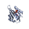
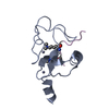
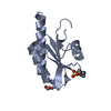
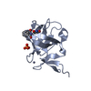
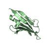
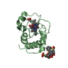
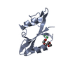
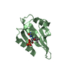
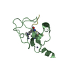
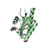
| Title | Crystal structure of the Phot-LOV1 domain from Chlamydomonas reinhardtii in illuminated state. Composite data set. | ||||||
 Components Components | putative blue light receptor | ||||||
 Keywords Keywords | ELECTRON TRANSPORT / phototropin / flavin | ||||||
| Function / homology |  Function and homology information Function and homology informationblue light signaling pathway / blue light photoreceptor activity / protein autophosphorylation / protein phosphorylation / non-specific serine/threonine protein kinase / protein serine kinase activity / protein serine/threonine kinase activity / ATP binding / nucleus / plasma membrane / cytoplasm Similarity search - Function | ||||||
| Biological species |   Chlamydomonas reinhardtii (plant) Chlamydomonas reinhardtii (plant) | ||||||
| Method |  X-RAY DIFFRACTION / SYNCHROTRON / MOLECULAR REPLACEMENT / Resolution: 2.8 Å X-RAY DIFFRACTION / SYNCHROTRON / MOLECULAR REPLACEMENT / Resolution: 2.8 Å | ||||||
 Authors Authors | Fedorov, R. / Schlichting, I. / Hartmann, E. / Domratcheva, T. / Fuhrmann, M. / Hegemann, P. | ||||||
 Citation Citation | Journal: Biophys.J. / Year: 2003 Title: Crystal structures and molecular mechanism of a light-induced signaling switch: The Phot-LOV1 domain from Chlamydomonas reinhardtii. Authors: Fedorov, R. / Schlichting, I. / Hartmann, E. / Domratcheva, T. / Fuhrmann, M. / Hegemann, P. | ||||||
| History |
|
- Structure visualization
| Structure viewer | Molecule: MolmilJmol/JSmol |
|---|
- Downloads & links
-Download
| PDBx/mmCIF format | 1n9o.cif.gz | 36.1 KB | Display | PDBx/mmCIF format |
|---|---|---|---|---|
| PDB format | pdb1n9o.ent.gz | 23.8 KB | Display | PDB format |
| PDBx/mmJSON format | 1n9o.json.gz | Tree view | PDBx/mmJSON format | |
| Others |  Other downloads Other downloads |
-Validation report
| Arichive directory | https://data.pdbj.org/pub/pdb/validation_reports/n9/1n9oftp://data.pdbj.org/pub/pdb/validation_reports/n9/1n9o | HTTPS FTP |
|---|
-Related structure data
| Related structure data | 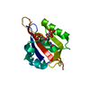 1n9lC 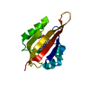 1n9nC 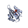 1g28S S: Starting model for refinement C: citing same article ( |
|---|---|
| Similar structure data |

-Links
 PDBj
PDBj


- Assembly
| Deposited unit | 
| ||||||||
|---|---|---|---|---|---|---|---|---|---|
| 1 |
| ||||||||
| 2 | 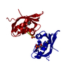
| ||||||||
| 3 | 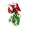
| ||||||||
| Unit cell |
| ||||||||
| Components on special symmetry positions |
|
-Components
| #1: Protein | Mass: 11989.646 Da / Num. of mol.: 1 / Fragment: residues 17-125 Source method: isolated from a genetically manipulated source Source: (gene. exp.) Chlamydomonas reinhardtii (plant) / Plasmid: pET16 / Species (production host): Escherichia coli / Production host:  |
|---|---|
| #2: Chemical | ChemComp-SO4 /   Mass: 96.063 Da / Num. of mol.: 1 / Source method: obtained synthetically / Formula: SO4 Mass: 96.063 Da / Num. of mol.: 1 / Source method: obtained synthetically / Formula: SO4 |
| #3: Chemical | ChemComp-FMN /   Mass: 456.344 Da / Num. of mol.: 1 / Source method: obtained synthetically / Formula: C17H21N4O9P Mass: 456.344 Da / Num. of mol.: 1 / Source method: obtained synthetically / Formula: C17H21N4O9P |
| #4: Water | ChemComp-HOH /  Mass: 18.015 Da / Num. of mol.: 26 / Source method: isolated from a natural source / Formula: H2O Mass: 18.015 Da / Num. of mol.: 26 / Source method: isolated from a natural source / Formula: H2O |
| Has protein modification | Y |
-Experimental details
-Experiment
| Experiment | Method: X-RAY DIFFRACTION / Number of used crystals: 2 |
|---|
- Sample preparation
| Crystal | Density Matthews: 3.89 Å3/Da / Density % sol: 68.13 % | |||||||||||||||||||||||||||||||||||||||||||||||||
|---|---|---|---|---|---|---|---|---|---|---|---|---|---|---|---|---|---|---|---|---|---|---|---|---|---|---|---|---|---|---|---|---|---|---|---|---|---|---|---|---|---|---|---|---|---|---|---|---|---|---|
| Crystal grow | Temperature: 293 K / Method: vapor diffusion, hanging drop / pH: 7.3 Details: HEPES, ammonium sulfate, PEG8000, pH 7.3, VAPOR DIFFUSION, HANGING DROP, temperature 293K | |||||||||||||||||||||||||||||||||||||||||||||||||
| Crystal grow | *PLUS Temperature: 20 ℃ / pH: 8 / Method: vapor diffusion, hanging drop | |||||||||||||||||||||||||||||||||||||||||||||||||
| Components of the solutions | *PLUS
|
-Data collection
| Diffraction |
| ||||||||||||||||||
|---|---|---|---|---|---|---|---|---|---|---|---|---|---|---|---|---|---|---|---|
| Diffraction source |
| ||||||||||||||||||
| Detector |
| ||||||||||||||||||
| Radiation |
| ||||||||||||||||||
| Radiation wavelength |
| ||||||||||||||||||
| Reflection | Resolution: 2.8→15 Å / Num. all: 5264 / Num. obs: 5009 / % possible obs: 95.2 % / Observed criterion σ(F): 2 / Observed criterion σ(I): 2 / Redundancy: 3.4 % / Biso Wilson estimate: 36.8 Å2 / Rsym value: 0.129 / Net I/σ(I): 7.5 | ||||||||||||||||||
| Reflection shell | Resolution: 2.8→2.9 Å / Mean I/σ(I) obs: 2.6 / Rsym value: 0.322 / % possible all: 82.5 | ||||||||||||||||||
| Reflection | *PLUS Num. all: 16983 / Rmerge(I) obs: 0.129 | ||||||||||||||||||
| Reflection shell | *PLUS % possible obs: 82.5 % / Rmerge(I) obs: 0.322 |

- Processing
| Software |
| ||||||||||||||||||||
|---|---|---|---|---|---|---|---|---|---|---|---|---|---|---|---|---|---|---|---|---|---|
| Refinement | Method to determine structure: MOLECULAR REPLACEMENT Starting model: PDB ENTRY 1G28 Resolution: 2.8→15 Å / Cross valid method: THROUGHOUT / σ(F): 0 / Stereochemistry target values: Engh & Huber
| ||||||||||||||||||||
| Refinement step | Cycle: LAST / Resolution: 2.8→15 Å
| ||||||||||||||||||||
| Refine LS restraints |
| ||||||||||||||||||||
| Refinement | *PLUS % reflection Rfree: 5 % / Rfactor Rwork: 0.25 | ||||||||||||||||||||
| Solvent computation | *PLUS | ||||||||||||||||||||
| Displacement parameters | *PLUS |
