 Movie
Movie Controller

[English] 日本語
 Yorodumi
- PDB-1g28: STRUCTURE OF A FLAVIN-BINDING DOMAIN, LOV2, FROM THE CHIMERIC PHY... -
+ Open data

- Basic information
| Entry | Database: PDB / ID: 1g28 | ||||||
|---|---|---|---|---|---|---|---|
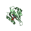
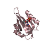
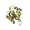
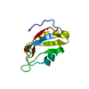
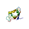
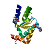
| Title | STRUCTURE OF A FLAVIN-BINDING DOMAIN, LOV2, FROM THE CHIMERIC PHYTOCHROME/PHOTOTROPIN PHOTORECEPTOR PHY3 | ||||||
 Components Components | PHY3 PROTEIN | ||||||
 Keywords Keywords | SIGNALING PROTEIN / ELECTRON TRANSPORT / phototropin / LOV / PAS fold / photoreceptor / flavoprotein / FMN-binding domain / alpha-beta structure | ||||||
| Function / homology |  Function and homology information Function and homology informationblue light photoreceptor activity / detection of visible light / non-specific serine/threonine protein kinase / protein serine/threonine kinase activity / regulation of DNA-templated transcription / DNA-templated transcription / ATP binding Similarity search - Function | ||||||
| Biological species |  Adiantum capillus-veneris (maidenhair fern) Adiantum capillus-veneris (maidenhair fern) | ||||||
| Method |  X-RAY DIFFRACTION / MOLECULAR REPLACEMENT / Resolution: 2.73 Å X-RAY DIFFRACTION / MOLECULAR REPLACEMENT / Resolution: 2.73 Å | ||||||
 Authors Authors | Crosson, S. / Moffat, K. | ||||||
 Citation Citation | Journal: Proc.Natl.Acad.Sci.USA / Year: 2001 Title: Structure of a flavin-binding plant photoreceptor domain: insights into light-mediated signal transduction Authors: Crosson, S. / Moffat, K. | ||||||
| History |
|
- Structure visualization
| Structure viewer | Molecule: MolmilJmol/JSmol |
|---|
- Downloads & links
-Download
| PDBx/mmCIF format | 1g28.cif.gz | 96.9 KB | Display | PDBx/mmCIF format |
|---|---|---|---|---|
| PDB format | pdb1g28.ent.gz | 74.5 KB | Display | PDB format |
| PDBx/mmJSON format | 1g28.json.gz | Tree view | PDBx/mmJSON format | |
| Others |  Other downloads Other downloads |
-Validation report
| Arichive directory | https://data.pdbj.org/pub/pdb/validation_reports/g2/1g28ftp://data.pdbj.org/pub/pdb/validation_reports/g2/1g28 | HTTPS FTP |
|---|
-Related structure data
| Similar structure data |
|---|


-Links
 PDBj
PDBj


- Assembly
| Deposited unit | 
| ||||||||||
|---|---|---|---|---|---|---|---|---|---|---|---|
| 1 | 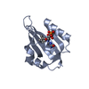
| ||||||||||
| 2 | 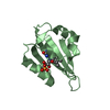
| ||||||||||
| 3 | 
| ||||||||||
| 4 | 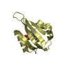
| ||||||||||
| Unit cell |
|
-Components
| #1: Protein | Mass: 12181.764 Da / Num. of mol.: 4 Fragment: FMN-BINDING DOMAIN OF CHIMERIC PHYTOCHROME/PHOTOTROPIN PHOTORECEPTOR, LOV3 Source method: isolated from a genetically manipulated source Source: (gene. exp.) Adiantum capillus-veneris (maidenhair fern)Gene: PHY3 / Plasmid: PCAL-NEK / Species (production host): Escherichia coli / Production host:  #2: Chemical | ChemComp-FMN /   Mass: 456.344 Da / Num. of mol.: 4 / Source method: obtained synthetically / Formula: C17H21N4O9P Mass: 456.344 Da / Num. of mol.: 4 / Source method: obtained synthetically / Formula: C17H21N4O9P#3: Water | ChemComp-HOH / |  Mass: 18.015 Da / Num. of mol.: 51 / Source method: isolated from a natural source / Formula: H2O Mass: 18.015 Da / Num. of mol.: 51 / Source method: isolated from a natural source / Formula: H2O |
|---|
-Experimental details
-Experiment
| Experiment | Method: X-RAY DIFFRACTION / Number of used crystals: 1 |
|---|
- Sample preparation
| Crystal | Density Matthews: 3.5 Å3/Da / Density % sol: 64.88 % | ||||||||||||||||||||
|---|---|---|---|---|---|---|---|---|---|---|---|---|---|---|---|---|---|---|---|---|---|
| Crystal grow | Temperature: 293 K / Method: vapor diffusion, hanging drop / pH: 8 Details: PEG 5000 MME, Tris, glycerol, pH 8.0, VAPOR DIFFUSION, HANGING DROP, temperature 293.0K | ||||||||||||||||||||
| Crystal grow | *PLUS Temperature: 20 ℃ / Method: vapor diffusion, sitting drop | ||||||||||||||||||||
| Components of the solutions | *PLUS
|
-Data collection
| Diffraction | Mean temperature: 298 K |
|---|---|
| Diffraction source | Source: ROTATING ANODE / Type: RIGAKU RU200 / Wavelength: 1.5418 Å |
| Detector | Type: RIGAKU RAXIS IIC / Detector: IMAGE PLATE / Date: Apr 24, 2000 / Details: mirrors |
| Radiation | Monochromator: Cu-K-alpha / Protocol: SINGLE WAVELENGTH / Monochromatic (M) / Laue (L): M / Scattering type: x-ray |
| Radiation wavelength | Wavelength: 1.5418 Å / Relative weight: 1 |
| Reflection | Resolution: 2.73→35.16 Å / Num. all: 17568 / Num. obs: 16249 / % possible obs: 92.5 % / Observed criterion σ(F): 0 / Observed criterion σ(I): -3 / Redundancy: 1.8 % / Biso Wilson estimate: 20.5 Å2 / Rmerge(I) obs: 0.115 / Net I/σ(I): 14.3 |
| Reflection shell | Resolution: 2.73→2.9 Å / Redundancy: 1.8 % / Rmerge(I) obs: 0.443 / % possible all: 86.1 |
| Reflection shell | *PLUS % possible obs: 86.1 % |
- Processing
| Software |
| |||||||||||||||||||||||||
|---|---|---|---|---|---|---|---|---|---|---|---|---|---|---|---|---|---|---|---|---|---|---|---|---|---|---|
| Refinement | Method to determine structure: MOLECULAR REPLACEMENT / Resolution: 2.73→35.16 Å / Rfactor Rfree error: 0.008 / Data cutoff high absF: 368490.28 / Data cutoff low absF: 0 / Isotropic thermal model: RESTRAINED / σ(F): 0 / σ(I): 0 / Stereochemistry target values: Engh & Huber
| |||||||||||||||||||||||||
| Solvent computation | Solvent model: FLAT MODEL / Bsol: 51.79 Å2 / ksol: 0.36 e/Å3 | |||||||||||||||||||||||||
| Displacement parameters | Biso mean: 34.2 Å2
| |||||||||||||||||||||||||
| Refine analyze |
| |||||||||||||||||||||||||
| Refinement step | Cycle: LAST / Resolution: 2.73→35.16 Å
| |||||||||||||||||||||||||
| Refine LS restraints |
| |||||||||||||||||||||||||
| Refine LS restraints NCS | NCS model details: CONSTR | |||||||||||||||||||||||||
| LS refinement shell | Resolution: 2.73→2.9 Å / Rfactor Rfree error: 0.032 / Total num. of bins used: 6
| |||||||||||||||||||||||||
| Software | *PLUS Name: CNS / Version: 1 / Classification: refinement | |||||||||||||||||||||||||
| Refine LS restraints | *PLUS
|
