 Movie
Movie Controller

[English] 日本語
 Yorodumi
- PDB-2eia: X-RAY CRYSTAL STRUCTURE OF EQUINE INFECTIOUS ANEMIA VIRUS (EIAV) ... -
+ Open data

- Basic information
| Entry | Database: PDB / ID: 2eia | ||||||
|---|---|---|---|---|---|---|---|
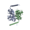
| Title | X-RAY CRYSTAL STRUCTURE OF EQUINE INFECTIOUS ANEMIA VIRUS (EIAV) CAPSID PROTEIN P26 | ||||||
 Components Components | EIAV CAPSID PROTEIN P26 | ||||||
 Keywords Keywords | VIRAL PROTEIN / VIRAL CAPSID EIAV / HIV / LENTIVIRUS | ||||||
| Function / homology |  Function and homology information Function and homology informationviral budding via host ESCRT complex / viral nucleocapsid / structural constituent of virion / nucleic acid binding / zinc ion binding Similarity search - Function | ||||||
| Biological species |  Equine infectious anemia virus Equine infectious anemia virus | ||||||
| Method |  X-RAY DIFFRACTION / SYNCHROTRON / MAD / Resolution: 2.7 Å X-RAY DIFFRACTION / SYNCHROTRON / MAD / Resolution: 2.7 Å | ||||||
 Authors Authors | Jin, Z. / Jin, L. / Peterson, D.L. / Lawson, C.L. | ||||||
 Citation Citation | Journal: J.Mol.Biol. / Year: 1999 Title: Model for lentivirus capsid core assembly based on crystal dimers of EIAV p26. Authors: Jin, Z. / Jin, L. / Peterson, D.L. / Lawson, C.L. #1: Journal: Biochim.Biophys.Acta / Year: 1997Title: Cloning, Expression, Purification, and Characterization of the Major Core Protein (P26) from Equine Infectious Anemia Virus Authors: Birkett, A.J. / Yelamos, B. / Rodriguez-Crespo, I. / Gavilanes, F. / Peterson, D.L. | ||||||
| History |
|
- Structure visualization
| Structure viewer | Molecule: MolmilJmol/JSmol |
|---|
- Downloads & links
-Download
| PDBx/mmCIF format | 2eia.cif.gz | 95.8 KB | Display | PDBx/mmCIF format |
|---|---|---|---|---|
| PDB format | pdb2eia.ent.gz | 74.1 KB | Display | PDB format |
| PDBx/mmJSON format | 2eia.json.gz | Tree view | PDBx/mmJSON format | |
| Others |  Other downloads Other downloads |
-Validation report
| Arichive directory | https://data.pdbj.org/pub/pdb/validation_reports/ei/2eiaftp://data.pdbj.org/pub/pdb/validation_reports/ei/2eia | HTTPS FTP |
|---|
-Related structure data


-Links
 PDBj
PDBj
- Assembly
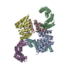
| Deposited unit | 
| ||||||||
|---|---|---|---|---|---|---|---|---|---|
| 1 | 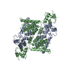
| ||||||||
| Unit cell |
| ||||||||
| Noncrystallographic symmetry (NCS) | NCS oper: (Code: given Matrix: (-0.544595, 0.821396, -0.169486), Vector: Details | THERE ARE TWO MONOMERS IN ONE ASYMMETRIC UNIT AND IN THIS MODEL. THE N-TERMINAL RESIDUES 1-16 AND C-TERMINAL RESIDUES 223-235 WERE NOT SEEN IN THE DENSITY MAPS. | |
-Components
| #1: Protein | Mass: 23386.543 Da / Num. of mol.: 2 Source method: isolated from a genetically manipulated source Source: (gene. exp.) Equine infectious anemia virus / Genus: Lentivirus / Gene: GAG / Gene (production host): GAG / Production host:  #2: Water | ChemComp-HOH / |  Mass: 18.015 Da / Num. of mol.: 188 / Source method: isolated from a natural source / Formula: H2O Mass: 18.015 Da / Num. of mol.: 188 / Source method: isolated from a natural source / Formula: H2OHas protein modification | Y | |
|---|
-Experimental details
-Experiment
| Experiment | Method: X-RAY DIFFRACTION / Number of used crystals: 1 |
|---|
- Sample preparation
| Crystal | Density Matthews: 2.23 Å3/Da / Density % sol: 44 % | ||||||||||||||||||||||||||||||
|---|---|---|---|---|---|---|---|---|---|---|---|---|---|---|---|---|---|---|---|---|---|---|---|---|---|---|---|---|---|---|---|
| Crystal grow | pH: 6.5 Details: 0.1M SODIUM CITRATE, 10% PEG 3350, 15% ISOPROPANOL, PH 6.5 | ||||||||||||||||||||||||||||||
| Crystal grow | *PLUS Method: vapor diffusion, hanging drop | ||||||||||||||||||||||||||||||
| Components of the solutions | *PLUS
|
-Data collection
| Diffraction | Mean temperature: 100 K | ||||||||||||
|---|---|---|---|---|---|---|---|---|---|---|---|---|---|
| Diffraction source | Source: SYNCHROTRON / Site: NSLS  / Beamline: X12C / Wavelength: 1.0333, 1.07168, 1.07202 / Beamline: X12C / Wavelength: 1.0333, 1.07168, 1.07202 | ||||||||||||
| Detector | Type: BRANDEIS / Detector: CCD / Date: Nov 11, 1997 / Details: MIRROR | ||||||||||||
| Radiation | Protocol: MAD / Monochromatic (M) / Laue (L): M / Scattering type: x-ray | ||||||||||||
| Radiation wavelength |
| ||||||||||||
| Reflection | Resolution: 2.7→47 Å / Num. obs: 13524 / % possible obs: 99.7 % / Observed criterion σ(I): 2 / Redundancy: 20 % / Biso Wilson estimate: 40.3 Å2 / Rmerge(I) obs: 0.063 / Net I/σ(I): 15 | ||||||||||||
| Reflection shell | Resolution: 2.7→2.8 Å / Redundancy: 12 % / Mean I/σ(I) obs: 3 / Rsym value: 0.283 / % possible all: 97.7 | ||||||||||||
| Reflection | *PLUS Num. measured all: 246510 / Rmerge(I) obs: 0.061 | ||||||||||||
| Reflection shell | *PLUS % possible obs: 97.7 % / Rmerge(I) obs: 0.326 |
- Processing
| Software |
| ||||||||||||||||||||||||||||||||||||||||||||||||||||||||||||
|---|---|---|---|---|---|---|---|---|---|---|---|---|---|---|---|---|---|---|---|---|---|---|---|---|---|---|---|---|---|---|---|---|---|---|---|---|---|---|---|---|---|---|---|---|---|---|---|---|---|---|---|---|---|---|---|---|---|---|---|---|---|
| Refinement | Method to determine structure: MAD / Resolution: 2.7→48 Å / Rfactor Rfree error: 0.008 / Data cutoff high rms absF: 274655 / Isotropic thermal model: RESTRAINED / Cross valid method: THROUGHOUT / σ(F): 2
| ||||||||||||||||||||||||||||||||||||||||||||||||||||||||||||
| Displacement parameters | Biso mean: 62.9 Å2
| ||||||||||||||||||||||||||||||||||||||||||||||||||||||||||||
| Refine analyze |
| ||||||||||||||||||||||||||||||||||||||||||||||||||||||||||||
| Refinement step | Cycle: LAST / Resolution: 2.7→48 Å
| ||||||||||||||||||||||||||||||||||||||||||||||||||||||||||||
| Refine LS restraints |
| ||||||||||||||||||||||||||||||||||||||||||||||||||||||||||||
| Refine LS restraints NCS | NCS model details: CONSTRAINED | ||||||||||||||||||||||||||||||||||||||||||||||||||||||||||||
| LS refinement shell | Resolution: 2.7→2.87 Å / Rfactor Rfree error: 0.034 / Total num. of bins used: 6
| ||||||||||||||||||||||||||||||||||||||||||||||||||||||||||||
| Xplor file |
| ||||||||||||||||||||||||||||||||||||||||||||||||||||||||||||
| Software | *PLUS Name: CNS / Version: 0.3 / Classification: refinement | ||||||||||||||||||||||||||||||||||||||||||||||||||||||||||||
| Refinement | *PLUS | ||||||||||||||||||||||||||||||||||||||||||||||||||||||||||||
| Solvent computation | *PLUS | ||||||||||||||||||||||||||||||||||||||||||||||||||||||||||||
| Displacement parameters | *PLUS | ||||||||||||||||||||||||||||||||||||||||||||||||||||||||||||
| Refine LS restraints | *PLUS
| ||||||||||||||||||||||||||||||||||||||||||||||||||||||||||||
| LS refinement shell | *PLUS Rfactor obs: 0.336 |
