National Institutes of Health/Office of the Director
P41 GM103403-10
United States
National Institutes of Health/National Institute of General Medical Sciences (NIH/NIGMS)
P30 GM124169
United States
Department of Energy (DOE, United States)
DE-AC02-05CH11231
United States
Citation
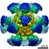
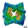
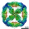
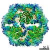
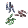
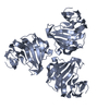
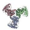
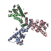
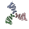
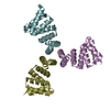
Journal: Elife / Year: 2020 Title: Tailored design of protein nanoparticle scaffolds for multivalent presentation of viral glycoprotein antigens. Authors: George Ueda / Aleksandar Antanasijevic / Jorge A Fallas / William Sheffler / Jeffrey Copps / Daniel Ellis / Geoffrey B Hutchinson / Adam Moyer / Anila Yasmeen / Yaroslav Tsybovsky / Young- ...Authors: George Ueda / Aleksandar Antanasijevic / Jorge A Fallas / William Sheffler / Jeffrey Copps / Daniel Ellis / Geoffrey B Hutchinson / Adam Moyer / Anila Yasmeen / Yaroslav Tsybovsky / Young-Jun Park / Matthew J Bick / Banumathi Sankaran / Rebecca A Gillespie / Philip Jm Brouwer / Peter H Zwart / David Veesler / Masaru Kanekiyo / Barney S Graham / Rogier W Sanders / John P Moore / Per Johan Klasse / Andrew B Ward / Neil P King / David Baker / Abstract: Multivalent presentation of viral glycoproteins can substantially increase the elicitation of antigen-specific antibodies. To enable a new generation of anti-viral vaccines, we designed self- ...Multivalent presentation of viral glycoproteins can substantially increase the elicitation of antigen-specific antibodies. To enable a new generation of anti-viral vaccines, we designed self-assembling protein nanoparticles with geometries tailored to present the ectodomains of influenza, HIV, and RSV viral glycoprotein trimers. We first designed trimers tailored for antigen fusion, featuring N-terminal helices positioned to match the C termini of the viral glycoproteins. Trimers that experimentally adopted their designed configurations were incorporated as components of tetrahedral, octahedral, and icosahedral nanoparticles, which were characterized by cryo-electron microscopy and assessed for their ability to present viral glycoproteins. Electron microscopy and antibody binding experiments demonstrated that the designed nanoparticles presented antigenically intact prefusion HIV-1 Env, influenza hemagglutinin, and RSV F trimers in the predicted geometries. This work demonstrates that antigen-displaying protein nanoparticles can be designed from scratch, and provides a systematic way to investigate the influence of antigen presentation geometry on the immune response to vaccination.
Mass: 13807.026 Da / Num. of mol.: 6 Source method: isolated from a genetically manipulated source Source: (gene. exp.) synthetic construct (others) / Production host: Escherichia coli (E. coli)
Method to determine structure: MOLECULAR REPLACEMENT Starting model: designed model from rosetta Resolution: 2.53→38.87 Å / Cor.coef. Fo:Fc: 0.948 / Cor.coef. Fo:Fc free: 0.912 / SU B: 22.749 / SU ML: 0.222 / Cross valid method: THROUGHOUT / ESU R: 0.641 / ESU R Free: 0.279 Details: U VALUES : WITH TLS ADDED HYDROGENS HAVE BEEN ADDED IN THE RIDING POSITIONS U VALUES : RESIDUAL ONLY
Rfactor
Num. reflection
% reflection
Selection details
Rfree
0.23166
1442
5.1 %
RANDOM
Rwork
0.18591
-
-
-
obs
0.18821
27066
99.86 %
-
Solvent computation
Ion probe radii: 0.7 Å / Shrinkage radii: 0.7 Å / VDW probe radii: 1 Å
Displacement parameters
Biso mean: 51.943 Å2
Baniso -1
Baniso -2
Baniso -3
1-
-0.59 Å2
0 Å2
0.79 Å2
2-
-
-0.2 Å2
-0 Å2
3-
-
-
0.28 Å2
Refinement step
Cycle: LAST / Resolution: 2.53→38.87 Å
Protein
Nucleic acid
Ligand
Solvent
Total
Num. atoms
5848
0
0
84
5932
Refine LS restraints
Refine-ID
Type
Dev ideal
Dev ideal target
Number
X-RAY DIFFRACTION
r_bond_refined_d
0.013
0.019
5987
X-RAY DIFFRACTION
r_bond_other_d
0.001
0.02
5144
X-RAY DIFFRACTION
r_angle_refined_deg
1.468
1.894
8110
X-RAY DIFFRACTION
r_angle_other_deg
1.225
2.933
11984
X-RAY DIFFRACTION
r_dihedral_angle_1_deg
5.105
5
706
X-RAY DIFFRACTION
r_dihedral_angle_2_deg
35.724
25.806
372
X-RAY DIFFRACTION
r_dihedral_angle_3_deg
18.139
15
978
X-RAY DIFFRACTION
r_dihedral_angle_4_deg
21.408
15
24
X-RAY DIFFRACTION
r_chiral_restr
0.098
0.2
774
X-RAY DIFFRACTION
r_gen_planes_refined
0.006
0.02
6925
X-RAY DIFFRACTION
r_gen_planes_other
0.001
0.02
1239
X-RAY DIFFRACTION
r_mcbond_it
4.407
2.392
2836
X-RAY DIFFRACTION
r_mcbond_other
4.407
2.391
2834
X-RAY DIFFRACTION
r_mcangle_it
5.998
3.58
3535
X-RAY DIFFRACTION
r_mcangle_other
5.997
3.58
3535
X-RAY DIFFRACTION
r_scbond_it
6.955
2.958
3151
X-RAY DIFFRACTION
r_scbond_other
6.954
2.958
3152
X-RAY DIFFRACTION
r_scangle_other
9.618
4.225
4575
X-RAY DIFFRACTION
r_long_range_B_refined
10.894
28.983
7170
X-RAY DIFFRACTION
r_long_range_B_other
10.896
28.928
7157
LS refinement shell
Resolution: 2.53→2.596 Å / Total num. of bins used: 20
Rfactor
Num. reflection
% reflection
Rfree
0.314
103
-
Rwork
0.273
2030
-
obs
-
-
99.91 %
Refinement TLS params.
Method: refined / Refine-ID: X-RAY DIFFRACTION
ID
L11 (°2)
L12 (°2)
L13 (°2)
L22 (°2)
L23 (°2)
L33 (°2)
S11 (Å °)
S12 (Å °)
S13 (Å °)
S21 (Å °)
S22 (Å °)
S23 (Å °)
S31 (Å °)
S32 (Å °)
S33 (Å °)
T11 (Å2)
T12 (Å2)
T13 (Å2)
T22 (Å2)
T23 (Å2)
T33 (Å2)
Origin x (Å)
Origin y (Å)
Origin z (Å)
1
3.1744
-0.3177
-1.1702
3.2054
-0.6563
4.2959
0.004
0.274
0.0458
-0.2581
-0.052
-0.2377
0.1539
0.0127
0.048
0.3054
-0.0081
-0.1126
0.0276
0.0122
0.0794
21.358
2.658
5.376
2
3.13
1.0105
0.9803
3.7581
0.7698
5.8867
0
-0.0852
-0.3005
0.1112
0.0601
-0.6756
-0.0298
0.9625
-0.0601
0.3167
-0.0003
-0.0647
0.1793
0.017
0.1502
39.029
11.527
35.834
3
2.0976
0.8446
-0.8787
3.2431
-1.0821
1.4953
-0.0104
0.0156
0.0842
0.0537
0.0923
0.2037
-0.1637
-0.1375
-0.0819
0.4175
0.0254
-0.082
0.0456
-0.0012
0.0332
14.443
-14.076
38.05
4
4.0213
-1.1822
0.7485
5.8281
-0.6909
3.3066
-0.1574
-0.1866
0.3699
0.2044
-0.0122
-0.8523
-0.0833
0.396
0.1695
0.3649
0.0067
-0.0898
0.0884
0.0002
0.1443
31.299
-28.69
28.043
5
2.9897
-0.0403
-0.5555
2.1197
1.0464
4.9866
0.0429
0.3153
0.1671
-0.2542
0.0965
-0.2172
-0.2109
0.1143
-0.1393
0.3218
-0.0012
-0.0724
0.0446
0.0162
0.1154
20.804
-37.56
-5.834
6
2.8279
-1.2019
-0.6099
3.8698
0.2713
0.5717
-0.0377
-0.0737
-0.1279
0.077
0.0733
0.203
0.0549
-0.053
-0.0356
0.2977
-0.0061
-0.1143
0.0155
0.0143
0.065
2.683
-11.891
11.036
Refinement TLS group
ID
Refine-ID
Refine TLS-ID
Auth asym-ID
Auth seq-ID
1
X-RAY DIFFRACTION
1
A
1 - 119
2
X-RAY DIFFRACTION
2
B
1 - 117
3
X-RAY DIFFRACTION
3
C
1 - 119
4
X-RAY DIFFRACTION
4
D
1 - 118
5
X-RAY DIFFRACTION
5
E
1 - 118
6
X-RAY DIFFRACTION
6
F
1 - 119
+
About Yorodumi
-
News
-
Feb 9, 2022. New format data for meta-information of EMDB entries
New format data for meta-information of EMDB entries
Version 3 of the EMDB header file is now the official format.
The previous official version 1.9 will be removed from the archive.
In the structure databanks used in Yorodumi, some data are registered as the other names, "COVID-19 virus" and "2019-nCoV". Here are the details of the virus and the list of structure data.
Jan 31, 2019. EMDB accession codes are about to change! (news from PDBe EMDB page)
EMDB accession codes are about to change! (news from PDBe EMDB page)
The allocation of 4 digits for EMDB accession codes will soon come to an end. Whilst these codes will remain in use, new EMDB accession codes will include an additional digit and will expand incrementally as the available range of codes is exhausted. The current 4-digit format prefixed with “EMD-” (i.e. EMD-XXXX) will advance to a 5-digit format (i.e. EMD-XXXXX), and so on. It is currently estimated that the 4-digit codes will be depleted around Spring 2019, at which point the 5-digit format will come into force.
The EM Navigator/Yorodumi systems omit the EMD- prefix.
Related info.:Q: What is EMD? / ID/Accession-code notation in Yorodumi/EM Navigator
Yorodumi is a browser for structure data from EMDB, PDB, SASBDB, etc.
This page is also the successor to EM Navigator detail page, and also detail information page/front-end page for Omokage search.
The word "yorodu" (or yorozu) is an old Japanese word meaning "ten thousand". "mi" (miru) is to see.
Related info.:EMDB / PDB / SASBDB / Comparison of 3 databanks / Yorodumi Search / Aug 31, 2016. New EM Navigator & Yorodumi / Yorodumi Papers / Jmol/JSmol / Function and homology information / Changes in new EM Navigator and Yorodumi
 Movie
Movie Controller

 Yorodumi
 Open data
 Basic information
 Components
 Keywords
 X-RAY DIFFRACTION /
  Authors
 United States, 5items
  Citation
 Structure visualization
 Molmil
 Downloads & links
 Other downloads


 PDBj
PDBj
 Assembly
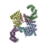

 Mass: 18.015 Da / Num. of mol.: 84 / Source method: isolated from a natural source / Formula: H2O
 Sample preparation
 Processing