 Movie
Movie Controller


+ Open data

- Basic information
| Entry | Database: EMDB / ID: EMD-21165 | |||||||||
|---|---|---|---|---|---|---|---|---|---|---|
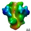
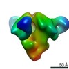
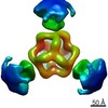
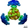
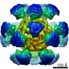
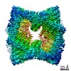
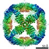
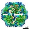
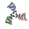
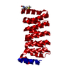
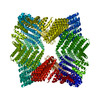
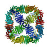
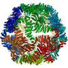
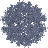
| Title | De novo designed octahedral nanoparticle O43_dn18 | |||||||||
 Map data Map data | O43_dn18 nanoparticle, Negative Stain EM map | |||||||||
 Sample Sample |
| |||||||||
| Biological species | synthetic construct (others) | |||||||||
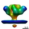
| Method | single particle reconstruction / negative staining / Resolution: 16.4 Å | |||||||||
 Authors Authors | Ward AB / Antanasijevic A | |||||||||
| Funding support |  United States, 1 items United States, 1 items
| |||||||||
 Citation Citation | Journal: Elife / Year: 2020 Title: Tailored design of protein nanoparticle scaffolds for multivalent presentation of viral glycoprotein antigens. Authors: George Ueda / Aleksandar Antanasijevic / Jorge A Fallas / William Sheffler / Jeffrey Copps / Daniel Ellis / Geoffrey B Hutchinson / Adam Moyer / Anila Yasmeen / Yaroslav Tsybovsky / Young- ...Authors: George Ueda / Aleksandar Antanasijevic / Jorge A Fallas / William Sheffler / Jeffrey Copps / Daniel Ellis / Geoffrey B Hutchinson / Adam Moyer / Anila Yasmeen / Yaroslav Tsybovsky / Young-Jun Park / Matthew J Bick / Banumathi Sankaran / Rebecca A Gillespie / Philip Jm Brouwer / Peter H Zwart / David Veesler / Masaru Kanekiyo / Barney S Graham / Rogier W Sanders / John P Moore / Per Johan Klasse / Andrew B Ward / Neil P King / David Baker /  Abstract: Multivalent presentation of viral glycoproteins can substantially increase the elicitation of antigen-specific antibodies. To enable a new generation of anti-viral vaccines, we designed self- ...Multivalent presentation of viral glycoproteins can substantially increase the elicitation of antigen-specific antibodies. To enable a new generation of anti-viral vaccines, we designed self-assembling protein nanoparticles with geometries tailored to present the ectodomains of influenza, HIV, and RSV viral glycoprotein trimers. We first designed trimers tailored for antigen fusion, featuring N-terminal helices positioned to match the C termini of the viral glycoproteins. Trimers that experimentally adopted their designed configurations were incorporated as components of tetrahedral, octahedral, and icosahedral nanoparticles, which were characterized by cryo-electron microscopy and assessed for their ability to present viral glycoproteins. Electron microscopy and antibody binding experiments demonstrated that the designed nanoparticles presented antigenically intact prefusion HIV-1 Env, influenza hemagglutinin, and RSV F trimers in the predicted geometries. This work demonstrates that antigen-displaying protein nanoparticles can be designed from scratch, and provides a systematic way to investigate the influence of antigen presentation geometry on the immune response to vaccination. | |||||||||
| History |
|
- Structure visualization
| Movie |
 Movie viewer Movie viewer |
|---|---|
| Structure viewer | EM map: SurfViewMolmilJmol/JSmol |
| Supplemental images |
 UCSF Chimera
- Downloads & links
-EMDB archive
| Map data | emd_21165.map.gz | 93.2 MB | EMDB map data format | |
|---|---|---|---|---|
| Header (meta data) | emd-21165-v30.xmlemd-21165.xml | 13.7 KB 13.7 KB | Display Display | EMDB header |
| Images | 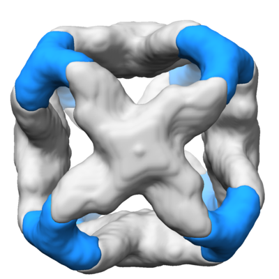 emd_21165.png emd_21165.png | 109.1 KB | ||
| Archive directory |  http://ftp.pdbj.org/pub/emdb/structures/EMD-21165ftp://ftp.pdbj.org/pub/emdb/structures/EMD-21165 http://ftp.pdbj.org/pub/emdb/structures/EMD-21165ftp://ftp.pdbj.org/pub/emdb/structures/EMD-21165 | HTTPS FTP |
-Related structure data


-Links
| EMDB pages | EMDB (EBI/PDBe) / EMDataResource |
|---|---|
| Related items in Molecule of the Month |

-Map
| File | Download / File: emd_21165.map.gz / Format: CCP4 / Size: 125 MB / Type: IMAGE STORED AS FLOATING POINT NUMBER (4 BYTES) | ||||||||||||||||||||||||||||||||||||||||||||||||||||||||||||||||||||
|---|---|---|---|---|---|---|---|---|---|---|---|---|---|---|---|---|---|---|---|---|---|---|---|---|---|---|---|---|---|---|---|---|---|---|---|---|---|---|---|---|---|---|---|---|---|---|---|---|---|---|---|---|---|---|---|---|---|---|---|---|---|---|---|---|---|---|---|---|---|
| Annotation | O43_dn18 nanoparticle, Negative Stain EM map | ||||||||||||||||||||||||||||||||||||||||||||||||||||||||||||||||||||
| Projections & slices | Image control
Images are generated by Spider. | ||||||||||||||||||||||||||||||||||||||||||||||||||||||||||||||||||||
| Voxel size | X=Y=Z: 2.05 Å | ||||||||||||||||||||||||||||||||||||||||||||||||||||||||||||||||||||
| Density |
| ||||||||||||||||||||||||||||||||||||||||||||||||||||||||||||||||||||
| Symmetry | Space group: 1 | ||||||||||||||||||||||||||||||||||||||||||||||||||||||||||||||||||||
| Details | EMDB XML:
CCP4 map header:
| ||||||||||||||||||||||||||||||||||||||||||||||||||||||||||||||||||||
 Z (Sec.)
 Y (Row.)
 X (Col.)


-Supplemental data
- Sample components
-Entire : De novo designed octahedral nanoparticle O43_dn18
| Entire | Name: De novo designed octahedral nanoparticle O43_dn18 |
|---|---|
| Components |
|
-Supramolecule #1: De novo designed octahedral nanoparticle O43_dn18
| Supramolecule | Name: De novo designed octahedral nanoparticle O43_dn18 / type: complex / ID: 1 / Parent: 0 / Macromolecule list: all / Details: Negative Stain EM Map |
|---|---|
| Source (natural) | Organism: synthetic construct (others) |
| Recombinant expression | Organism:  |
| Molecular weight | Theoretical: 880 KDa |
-Macromolecule #1: O43_dn18B
| Macromolecule | Name: O43_dn18B / type: protein_or_peptide / ID: 1 / Enantiomer: LEVO |
|---|---|
| Source (natural) | Organism: synthetic construct (others) |
| Recombinant expression | Organism: |
| Sequence | String: MGEEAELAYL LGELAYKLGE YRIAIRAYRI ALKRDPNNAE AWYNLGNAYY KQGDYDEAIE YYQKALELDP NNAEAWYNLG NAYYKQGDYD EAIEYYQKAL ELDPSNLDAA VNLGAATMLT S |
-Macromolecule #2: O43_dn18A
| Macromolecule | Name: O43_dn18A / type: protein_or_peptide / ID: 2 / Enantiomer: LEVO |
|---|---|
| Source (natural) | Organism: synthetic construct (others) |
| Recombinant expression | Organism: |
| Sequence | String: MDRCEELARR IAEVVERAKR AGTSEDEIAE SVARVISLVI RALKLSGSSY EVICECVARI VAEIVEALKR SGTSAVEIAK IVARVISEVI RTLKESGSSY EVICECVARI VAEIVEALKR SGTSAAIIAL IVALVISEVI RTLKESGSSF EVILECVIRI VLEIIEALKR ...String: MDRCEELARR IAEVVERAKR AGTSEDEIAE SVARVISLVI RALKLSGSSY EVICECVARI VAEIVEALKR SGTSAVEIAK IVARVISEVI RTLKESGSSY EVICECVARI VAEIVEALKR SGTSAAIIAL IVALVISEVI RTLKESGSSF EVILECVIRI VLEIIEALKR SGTSEQDVML IVMAVLLVVL ATLQLSGSGG WLEHHHHHH |
-Experimental details
-Structure determination
| Method | negative staining |
|---|---|
 Processing Processing | single particle reconstruction |
| Aggregation state | particle |
-Sample preparation
| Concentration | 0.05 mg/mL | |||||||||
|---|---|---|---|---|---|---|---|---|---|---|
| Buffer | pH: 7.4 Component:
Details: TBS buffer, pH 7.4 | |||||||||
| Staining | Type: NEGATIVE / Material: Uranyl Formate Details: Sample diluted to 0.05 mg/mL. 3 uL was applied onto the grid, blotted off, and then stained with 2% uranyl formate for 60 seconds. | |||||||||
| Grid | Material: COPPER / Mesh: 400 / Support film - Material: CARBON / Support film - topology: CONTINUOUS | |||||||||
| Details | O43_dn18 nanoparticle was purified by SEC, diluted to 0.05mg/ml and loaded onto a grid. |
- Electron microscopy
| Microscope | FEI TECNAI SPIRIT |
|---|---|
| Image recording | Film or detector model: TVIPS TEMCAM-F416 (4k x 4k) / Average electron dose: 25.0 e/Å2 |
| Electron beam | Acceleration voltage: 120 kV / Electron source: LAB6 |
| Electron optics | C2 aperture diameter: 100.0 µm / Illumination mode: FLOOD BEAM / Imaging mode: BRIGHT FIELD / Cs: 2.7 mm / Nominal defocus max: 1.5 µm / Nominal defocus min: 1.5 µm |
| Experimental equipment |  Model: Tecnai Spirit / Image courtesy: FEI Company |
