[English] 日本語
Yorodumi
- PDB-6veh: Computationally designed C3-symmetric homotrimer from HEAT repeat... -

+
Open data


ID or keywords:

Loading...

-
Basic information

Entry
Database: PDB / ID: 6veh
TitleComputationally designed C3-symmetric homotrimer from HEAT repeat protein
ComponentsHEAT repeat domain-containing protein
KeywordsDE NOVO PROTEIN / designed protein / vaccine
Biological speciessynthetic construct (others)
MethodX-RAY DIFFRACTION / SYNCHROTRON / MOLECULAR REPLACEMENT / molecular replacement / Resolution: 2.303 Å
AuthorsBick, M.J. / Ueda, G. / Baker, D.
Funding support United States, 4items
OrganizationGrant numberCountry
Department of Energy (DOE, United States)DE-AC02-05CH11231 United States
National Institutes of Health/National Institute of General Medical Sciences (NIH/NIGMS)P30GM124169 United States
National Institutes of Health/National Institute of General Medical Sciences (NIH/NIGMS)S10OD018483 United States
Howard Hughes Medical Institute (HHMI) United States
Citation
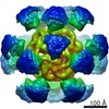
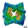
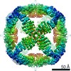
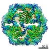
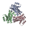
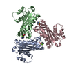
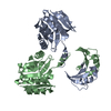
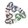
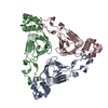
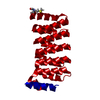
Journal: Elife / Year: 2020
Title: Tailored design of protein nanoparticle scaffolds for multivalent presentation of viral glycoprotein antigens.
Authors: George Ueda / Aleksandar Antanasijevic / Jorge A Fallas / William Sheffler / Jeffrey Copps / Daniel Ellis / Geoffrey B Hutchinson / Adam Moyer / Anila Yasmeen / Yaroslav Tsybovsky / Young- ...Authors: George Ueda / Aleksandar Antanasijevic / Jorge A Fallas / William Sheffler / Jeffrey Copps / Daniel Ellis / Geoffrey B Hutchinson / Adam Moyer / Anila Yasmeen / Yaroslav Tsybovsky / Young-Jun Park / Matthew J Bick / Banumathi Sankaran / Rebecca A Gillespie / Philip Jm Brouwer / Peter H Zwart / David Veesler / Masaru Kanekiyo / Barney S Graham / Rogier W Sanders / John P Moore / Per Johan Klasse / Andrew B Ward / Neil P King / David Baker /
Abstract: Multivalent presentation of viral glycoproteins can substantially increase the elicitation of antigen-specific antibodies. To enable a new generation of anti-viral vaccines, we designed self- ...Multivalent presentation of viral glycoproteins can substantially increase the elicitation of antigen-specific antibodies. To enable a new generation of anti-viral vaccines, we designed self-assembling protein nanoparticles with geometries tailored to present the ectodomains of influenza, HIV, and RSV viral glycoprotein trimers. We first designed trimers tailored for antigen fusion, featuring N-terminal helices positioned to match the C termini of the viral glycoproteins. Trimers that experimentally adopted their designed configurations were incorporated as components of tetrahedral, octahedral, and icosahedral nanoparticles, which were characterized by cryo-electron microscopy and assessed for their ability to present viral glycoproteins. Electron microscopy and antibody binding experiments demonstrated that the designed nanoparticles presented antigenically intact prefusion HIV-1 Env, influenza hemagglutinin, and RSV F trimers in the predicted geometries. This work demonstrates that antigen-displaying protein nanoparticles can be designed from scratch, and provides a systematic way to investigate the influence of antigen presentation geometry on the immune response to vaccination.
History
DepositionJan 2, 2020Deposition site: RCSB / Processing site: RCSB
Revision 1.0Aug 5, 2020Provider: repository / Type: Initial release
Revision 1.1Aug 19, 2020Group: Database references / Category: citation / citation_author
Item: _citation.country / _citation.journal_abbrev ..._citation.country / _citation.journal_abbrev / _citation.journal_id_CSD / _citation.journal_id_ISSN / _citation.journal_volume / _citation.pdbx_database_id_DOI / _citation.pdbx_database_id_PubMed / _citation.title / _citation.year
Revision 1.2Apr 3, 2024Group: Data collection / Database references / Refinement description
Category: chem_comp_atom / chem_comp_bond ...chem_comp_atom / chem_comp_bond / database_2 / pdbx_initial_refinement_model
Item: _database_2.pdbx_DOI / _database_2.pdbx_database_accession

-
Structure visualization

Structure viewerMolecule:
MolmilJmol/JSmol

Downloads & links

-
Assembly

Deposited unit
A: HEAT repeat domain-containing protein


Theoretical massNumber of molelcules
Total (without water)21,0221
Polymers21,0221
Non-polymers00
Water45025
1
A: HEAT repeat domain-containing protein

A: HEAT repeat domain-containing protein

A: HEAT repeat domain-containing protein


Theoretical massNumber of molelcules
Total (without water)63,0673
Polymers63,0673
Non-polymers00
Water543
TypeNameSymmetry operationNumber
identity operation1_555x,y,z1
crystal symmetry operation2_655-y+1,x-y,z1
crystal symmetry operation3_665-x+y+1,-x+1,z1
Buried area4540 Å2
ΔGint-61 kcal/mol
Surface area22780 Å2
MethodPISA
Unit cell
Length a, b, c (Å)88.182, 88.182, 65.244
Angle α, β, γ (deg.)90.000, 90.000, 120.000
Int Tables number146
Space group name H-MH3

-
Components

#1: Protein HEAT repeat domain-containing protein


Mass: 21022.387 Da / Num. of mol.: 1
Source method: isolated from a genetically manipulated source
Source: (gene. exp.) synthetic construct (others) / Production host: Escherichia coli (E. coli)
#2: Water ChemComp-HOH / water / Water


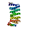
Mass: 18.015 Da / Num. of mol.: 25 / Source method: isolated from a natural source / Formula: H2O
Has ligand of interestN

-
Experimental details

-
Experiment

ExperimentMethod: X-RAY DIFFRACTION / Number of used crystals: 1

-
Sample preparation

CrystalDensity Matthews: 2.32 Å3/Da / Density % sol: 47.03 %
Crystal growTemperature: 291.15 K / Method: vapor diffusion, sitting drop
Details: 1mM DL-glutamic acid monohydrate, 100mM DL-alanine, 100mM glycine, 100mM DL-lysine monohydrochloride, 100mM DL-serine, 100mM Tris (base), 100mM BICINE, 20% (v/v) ethylene glycol, 10 % (w/v) PEG 8000, pH8.5

-
Data collection

DiffractionMean temperature: 100 K / Ambient temp details: liquid nitrogen stream / Serial crystal experiment: N
Diffraction sourceSource: SYNCHROTRON / Site: ALS / Beamline: 5.0.1 / Wavelength: 0.977408 Å
DetectorType: ADSC QUANTUM 315r / Detector: CCD / Date: Feb 10, 2016
RadiationProtocol: SINGLE WAVELENGTH / Monochromatic (M) / Laue (L): M / Scattering type: x-ray
Radiation wavelengthWavelength: 0.977408 Å / Relative weight: 1
ReflectionResolution: 2.3→100 Å / Num. obs: 8361 / % possible obs: 100 % / Redundancy: 5.6 % / Biso Wilson estimate: 40.43 Å2 / Rmerge(I) obs: 0.119 / Rpim(I) all: 0.055 / Rrim(I) all: 0.131 / Χ2: 0.944 / Net I/σ(I): 6.2 / Num. measured all: 46905
Reflection shell

Diffraction-ID: 1

Resolution (Å)Redundancy (%)Rmerge(I) obsNum. unique obsCC1/2Rpim(I) allRrim(I) allΧ2% possible all
2.3-2.3451.1983880.6190.5981.3410.714100
2.34-2.385.20.9484540.5860.4581.0530.673100
2.38-2.435.40.8454100.6530.40.9350.759100
2.43-2.485.60.8354230.780.3890.9210.715100
2.48-2.535.50.6174030.8050.290.6831.248100
2.53-2.595.70.6744240.7930.3120.7430.769100
2.59-2.665.70.5464180.8780.2530.6030.86100
2.66-2.735.70.4114120.9040.190.4530.95100
2.73-2.815.70.384300.9160.1750.4181.023100
2.81-2.95.70.2994270.9520.1380.331.003100
2.9-35.70.2793860.950.1280.3070.958100
3-3.125.70.2554310.9510.1170.281.03100
3.12-3.265.70.2284280.9650.1060.2511.004100
3.26-3.445.60.1524080.9730.070.1670.968100
3.44-3.655.70.144350.9870.0640.1540.985100
3.65-3.935.70.1244170.9840.0570.1371.005100
3.93-4.335.60.0884090.9930.0410.0970.993100
4.33-4.965.50.0674230.9950.0320.0741.059100
4.96-6.245.90.0594160.9970.0270.0651.039100
6.24-1005.80.0334190.9990.0150.0361.07799.8

-
Phasing

PhasingMethod: molecular replacement

-
Processing

Software
NameVersionClassification
HKL-2000714ndata reduction
HKL-2000714ndata scaling
PHASER2.8.2phasing
PHENIXdev_3112refinement
PDB_EXTRACT3.25data extraction
RefinementMethod to determine structure: MOLECULAR REPLACEMENT
Starting model: Computational design model

Resolution: 2.303→44.091 Å / SU ML: 0.25 / Cross valid method: THROUGHOUT / σ(F): 0.15 / Phase error: 25.64
RfactorNum. reflection% reflection
Rfree0.2261 756 9.88 %
Rwork0.2021 --
obs0.2045 7654 91.52 %
Solvent computationShrinkage radii: 0.9 Å / VDW probe radii: 1.11 Å
Displacement parametersBiso max: 121.21 Å2 / Biso mean: 53.4257 Å2 / Biso min: 26.15 Å2
Refinement stepCycle: final / Resolution: 2.303→44.091 Å
ProteinNucleic acidLigandSolventTotal
Num. atoms1249 0 0 25 1274
Biso mean---50.8 -
Num. residues----180
LS refinement shell

Refine-ID: X-RAY DIFFRACTION / Rfactor Rfree error: 0

Resolution (Å)Rfactor RfreeNum. reflection RfreeRfactor RworkNum. reflection Rwork% reflection obs (%)
2.3035-2.48130.271280.2417121080
2.4813-2.7310.30521510.2316132189
2.731-3.12610.24671540.2119142994
3.1261-3.93820.20641570.1929144795
3.9382-44.0910.20851660.1919149199

+
About Yorodumi

-
News

-
Feb 9, 2022. New format data for meta-information of EMDB entries

New format data for meta-information of EMDB entries

  • Version 3 of the EMDB header file is now the official format.
  • The previous official version 1.9 will be removed from the archive.

Related info.:EMDB header

External links:wwPDB to switch to version 3 of the EMDB data model

-
Aug 12, 2020. Covid-19 info

Covid-19 info

URL: https://pdbj.org/emnavi/covid19.php

New page: Covid-19 featured information page in EM Navigator.

Related info.:Covid-19 info / Mar 5, 2020. Novel coronavirus structure data

+
Mar 5, 2020. Novel coronavirus structure data

Novel coronavirus structure data

Related info.:Yorodumi Speices / Aug 12, 2020. Covid-19 info

External links:COVID-19 featured content - PDBj / Molecule of the Month (242):Coronavirus Proteases

+
Jan 31, 2019. EMDB accession codes are about to change! (news from PDBe EMDB page)

EMDB accession codes are about to change! (news from PDBe EMDB page)

  • The allocation of 4 digits for EMDB accession codes will soon come to an end. Whilst these codes will remain in use, new EMDB accession codes will include an additional digit and will expand incrementally as the available range of codes is exhausted. The current 4-digit format prefixed with “EMD-” (i.e. EMD-XXXX) will advance to a 5-digit format (i.e. EMD-XXXXX), and so on. It is currently estimated that the 4-digit codes will be depleted around Spring 2019, at which point the 5-digit format will come into force.
  • The EM Navigator/Yorodumi systems omit the EMD- prefix.

Related info.:Q: What is EMD? / ID/Accession-code notation in Yorodumi/EM Navigator

External links:EMDB Accession Codes are Changing Soon! / Contact to PDBj

+
Jul 12, 2017. Major update of PDB

Major update of PDB

  • wwPDB released updated PDB data conforming to the new PDBx/mmCIF dictionary.
  • This is a major update changing the version number from 4 to 5, and with Remediation, in which all the entries are updated.
  • In this update, many items about electron microscopy experimental information are reorganized (e.g. em_software).
  • Now, EM Navigator and Yorodumi are based on the updated data.

External links:wwPDB Remediation / Enriched Model Files Conforming to OneDep Data Standards Now Available in the PDB FTP Archive

-
Yorodumi

Thousand views of thousand structures

  • Yorodumi is a browser for structure data from EMDB, PDB, SASBDB, etc.
  • This page is also the successor to EM Navigator detail page, and also detail information page/front-end page for Omokage search.
  • The word "yorodu" (or yorozu) is an old Japanese word meaning "ten thousand". "mi" (miru) is to see.

Related info.:EMDB / PDB / SASBDB / Comparison of 3 databanks / Yorodumi Search / Aug 31, 2016. New EM Navigator & Yorodumi / Yorodumi Papers / Jmol/JSmol / Function and homology information / Changes in new EM Navigator and Yorodumi

Read more