 Movie
Movie Controller

[English] 日本語
 Yorodumi
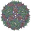
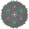
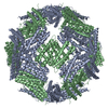
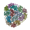
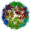
- PDB-2vtu: crystal structure of bacteriophage MS2 covalent coat protein dimer -
+ Open data

- Basic information
| Entry | Database: PDB / ID: 2vtu | ||||||
|---|---|---|---|---|---|---|---|
| Title | crystal structure of bacteriophage MS2 covalent coat protein dimer | ||||||
 Components Components | MS2 COAT PROTEIN | ||||||
 Keywords Keywords | VIRUS / MS2 / DIMER / VIRION / OCTAHEDRON / RNA-BINDING / COAT PROTEIN / CAPSID PROTEIN | ||||||
| Function / homology | negative regulation of viral translation / Levivirus coat protein / Levivirus coat protein / Bacteriophage RNA-type, capsid / T=3 icosahedral viral capsid / structural molecule activity / RNA binding / identical protein binding / Capsid protein Function and homology information Function and homology information | ||||||
| Biological species |  ENTEROBACTERIO PHAGE MS2 (virus) ENTEROBACTERIO PHAGE MS2 (virus) | ||||||
| Method |  X-RAY DIFFRACTION / SYNCHROTRON / MOLECULAR REPLACEMENT / Resolution: 3.5 Å X-RAY DIFFRACTION / SYNCHROTRON / MOLECULAR REPLACEMENT / Resolution: 3.5 Å | ||||||
 Authors Authors | Plevka, P. / Tars, K. / Liljas, L. | ||||||
 Citation Citation | Journal: Protein Sci. / Year: 2008 Title: Crystal Packing of a Bacteriophage MS2 Coat Protein Mutant Corresponds to Octahedral Particles. Authors: Plevka, P. / Tars, K. / Liljas, L. | ||||||
| History |
| ||||||
| Remark 650 | HELIX DETERMINATION METHOD: AUTHOR PROVIDED. | ||||||
| Remark 700 | SHEET THE SHEET STRUCTURE OF THIS MOLECULE IS BIFURCATED. IN ORDER TO REPRESENT THIS FEATURE IN ... SHEET THE SHEET STRUCTURE OF THIS MOLECULE IS BIFURCATED. IN ORDER TO REPRESENT THIS FEATURE IN THE SHEET RECORDS BELOW, TWO SHEETS ARE DEFINED. |
- Structure visualization
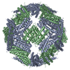
| Structure viewer | Molecule: MolmilJmol/JSmol |
|---|
- Downloads & links
-Download
| PDBx/mmCIF format | 2vtu.cif.gz | 175.9 KB | Display | PDBx/mmCIF format |
|---|---|---|---|---|
| PDB format | pdb2vtu.ent.gz | 148.2 KB | Display | PDB format |
| PDBx/mmJSON format | 2vtu.json.gz | Tree view | PDBx/mmJSON format | |
| Others |  Other downloads Other downloads |
-Validation report
| Arichive directory | https://data.pdbj.org/pub/pdb/validation_reports/vt/2vtuftp://data.pdbj.org/pub/pdb/validation_reports/vt/2vtu | HTTPS FTP |
|---|
-Related structure data
| Related structure data | 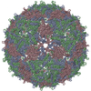 2ms2S S: Starting model for refinement |
|---|---|
| Similar structure data |


-Links
 PDBj
PDBj
- Assembly
| Deposited unit | 
| ||||||||
|---|---|---|---|---|---|---|---|---|---|
| 1 | x 24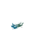
| ||||||||
| 2 |
| ||||||||
| 3 | 
| ||||||||
| Unit cell |
| ||||||||
| Number of models | 2 | ||||||||
| Symmetry | Point symmetry: (Schoenflies symbol: O (octahedral)) | ||||||||
| Noncrystallographic symmetry (NCS) | NCS oper: (Code: given / Matrix: (1), |
-Components
| #1: Protein | Mass: 27371.877 Da / Num. of mol.: 2 / Fragment: RESIDUES 2-130,2 AND 4-130 Source method: isolated from a genetically manipulated source Details: EACH CHAIN COMPRISES A REPEAT UNIT CONTAINING RESIDUES 2-130 (PDB RESIDUES 1-129), RESIDUE 2 (PDB RESIDUE 130) AND 4-130 (PDB RESIDUES 131-257) Source: (gene. exp.) ENTEROBACTERIO PHAGE MS2 (virus) / Plasmid: PBAD / Production host:  |
|---|
-Experimental details
-Experiment
| Experiment | Method: X-RAY DIFFRACTION / Number of used crystals: 1 |
|---|
- Sample preparation
| Crystal | Density Matthews: 2.7 Å3/Da / Density % sol: 47 % / Description: NONE |
|---|---|
| Crystal grow | pH: 7.5 Details: 0.32M NA2HPO4, 0.08M NAH2PO4 AND 5% PEG 8000, pH 7.5 |
-Data collection
| Diffraction | Mean temperature: 100 K |
|---|---|
| Diffraction source | Source: SYNCHROTRON / Site: ESRF  / Beamline: ID14-4 / Wavelength: 0.934 / Beamline: ID14-4 / Wavelength: 0.934 |
| Detector | Type: ADSC CCD / Detector: CCD / Date: May 19, 2005 |
| Radiation | Protocol: SINGLE WAVELENGTH / Monochromatic (M) / Laue (L): M / Scattering type: x-ray |
| Radiation wavelength | Wavelength: 0.934 Å / Relative weight: 1 |
| Reflection | Resolution: 3.5→45 Å / Num. obs: 6079 / % possible obs: 97.8 % / Observed criterion σ(I): 2 / Redundancy: 5.6 % / Rmerge(I) obs: 0.15 / Net I/σ(I): 17.3 |
| Reflection shell | Resolution: 3.5→3.63 Å / Redundancy: 5.7 % / Rmerge(I) obs: 0.36 / Mean I/σ(I) obs: 6.7 / % possible all: 99.2 |
- Processing
| Software |
| ||||||||||||||||||||||||||||||||||||||||||||||||||||||||||||
|---|---|---|---|---|---|---|---|---|---|---|---|---|---|---|---|---|---|---|---|---|---|---|---|---|---|---|---|---|---|---|---|---|---|---|---|---|---|---|---|---|---|---|---|---|---|---|---|---|---|---|---|---|---|---|---|---|---|---|---|---|---|
| Refinement | Method to determine structure: MOLECULAR REPLACEMENT Starting model: PDB ENTRY 2MS2 Resolution: 3.5→45 Å / Rfactor Rfree error: 0.083 / Data cutoff high absF: 10000 / Cross valid method: THROUGHOUT / σ(F): 0 Stereochemistry target values: MAXIMUM LIKELIHOOD TARGET USING AMPLITUDES Details: THE DIFFRACTION DATA REPRESENTS AN AVERAGE OF TWO ORIENTATIONS DEMONSTRATED BY THE TWO MODELS. CHAIN J IN MODEL 1 AND 2 ARE APPROXIMATELY 180 DEGREES ROTATED TO EACH OTHER. THE SUBUNIT L HAS ...Details: THE DIFFRACTION DATA REPRESENTS AN AVERAGE OF TWO ORIENTATIONS DEMONSTRATED BY THE TWO MODELS. CHAIN J IN MODEL 1 AND 2 ARE APPROXIMATELY 180 DEGREES ROTATED TO EACH OTHER. THE SUBUNIT L HAS 0.25 OCCUPANCY, BECAUSE TWOFOLD CRYSTALLOGRAPHIC AXIS PUTS 180 DEGREE ROTATED COPY OF THE L SUBUNIT ON TOP OF ITSELF.
| ||||||||||||||||||||||||||||||||||||||||||||||||||||||||||||
| Solvent computation | Solvent model: FLAT MODEL / Bsol: 15.2947 Å2 / ksol: 0.984345 e/Å3 | ||||||||||||||||||||||||||||||||||||||||||||||||||||||||||||
| Displacement parameters | Biso mean: 15.3 Å2
| ||||||||||||||||||||||||||||||||||||||||||||||||||||||||||||
| Refine analyze |
| ||||||||||||||||||||||||||||||||||||||||||||||||||||||||||||
| Refinement step | Cycle: LAST / Resolution: 3.5→45 Å
| ||||||||||||||||||||||||||||||||||||||||||||||||||||||||||||
| Refine LS restraints |
| ||||||||||||||||||||||||||||||||||||||||||||||||||||||||||||
| LS refinement shell | Resolution: 3.5→3.72 Å / Rfactor Rfree error: 0.083 / Total num. of bins used: 6
| ||||||||||||||||||||||||||||||||||||||||||||||||||||||||||||
| Xplor file | Serial no: 1 / Param file: CNS_TOPPAR_PROTEIN_REP.PARAM / Topol file: CNS_TOPPAR_PROTEIN.TOP |
