 Movie
Movie Controller

[English] 日本語
 Yorodumi
- PDB-2vn5: The Clostridium cellulolyticum dockerin displays a dual binding m... -
+ Open data

- Basic information
| Entry | Database: PDB / ID: 2vn5 | ||||||
|---|---|---|---|---|---|---|---|
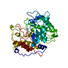
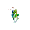
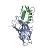
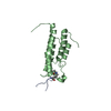
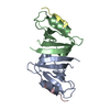
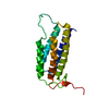
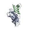
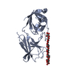
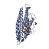
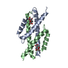
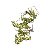
| Title | The Clostridium cellulolyticum dockerin displays a dual binding mode for its cohesin partner | ||||||
 Components Components |
| ||||||
 Keywords Keywords | CELL ADHESION / CARBOHYDRATE METABOLISM / POLYSACCHARIDE DEGRADATION / COHESIN / DOCKERIN / HYDROLASE / CELLULOSOME / GLYCOSIDASE / CELLULOSE DEGRADATION | ||||||
| Function / homology |  Function and homology information Function and homology informationcellulose binding / cellulase / cellulase activity / beta-glucosidase activity / cellulose catabolic process / cell surface / extracellular region / metal ion binding Similarity search - Function | ||||||
| Biological species |  CLOSTRIDIUM CELLULOLYTICUM (bacteria) CLOSTRIDIUM CELLULOLYTICUM (bacteria) | ||||||
| Method |  X-RAY DIFFRACTION / SYNCHROTRON / MOLECULAR REPLACEMENT / Resolution: 1.9 Å X-RAY DIFFRACTION / SYNCHROTRON / MOLECULAR REPLACEMENT / Resolution: 1.9 Å | ||||||
 Authors Authors | Pinheiro, B.A. / Prates, J.A.M. / Proctor, M.R. / Gilbert, H.J. / Davies, G.J. / Money, V.A. / Martinez-Fleites, C. / Bayer, E.A. / Fontes, C.M.G.A. / Fierobe, H.P. | ||||||
 Citation Citation | Journal: J.Biol.Chem. / Year: 2008 Title: The Clostridium Cellulolyticum Dockerin Displays a Dual Binding Mode for its Cohesin Partner. Authors: Pinheiro, B.A. / Proctor, M.R. / Martinez-Fleites, C. / Prates, J.A.M. / Money, V.A. / Davies, G.J. / Bayer, E.A. / Fontes, C.M.G.A. / Fierobe, H.P. / Gilbert, H.J. | ||||||
| History |
| ||||||
| Remark 700 | SHEET THE SHEET STRUCTURE OF THIS MOLECULE IS BIFURCATED. IN ORDER TO REPRESENT THIS FEATURE IN ... SHEET THE SHEET STRUCTURE OF THIS MOLECULE IS BIFURCATED. IN ORDER TO REPRESENT THIS FEATURE IN THE SHEET RECORDS BELOW, TWO SHEETS ARE DEFINED. |
- Structure visualization
| Structure viewer | Molecule: MolmilJmol/JSmol |
|---|
- Downloads & links
-Download
| PDBx/mmCIF format | 2vn5.cif.gz | 93.2 KB | Display | PDBx/mmCIF format |
|---|---|---|---|---|
| PDB format | pdb2vn5.ent.gz | 71 KB | Display | PDB format |
| PDBx/mmJSON format | 2vn5.json.gz | Tree view | PDBx/mmJSON format | |
| Others |  Other downloads Other downloads |
-Validation report
| Arichive directory | https://data.pdbj.org/pub/pdb/validation_reports/vn/2vn5ftp://data.pdbj.org/pub/pdb/validation_reports/vn/2vn5 | HTTPS FTP |
|---|
-Related structure data
| Related structure data |  2vn6C  1ohzS C: citing same article ( S: Starting model for refinement |
|---|---|
| Similar structure data |


-Links
 PDBj
PDBj
- Assembly
| Deposited unit | 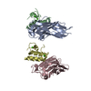
| ||||||||
|---|---|---|---|---|---|---|---|---|---|
| 1 | 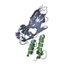
| ||||||||
| 2 | 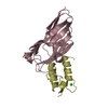
| ||||||||
| Unit cell |
|
-Components
| #1: Protein | Mass: 15557.599 Da / Num. of mol.: 2 / Fragment: RESIDUES 277-427 Source method: isolated from a genetically manipulated source Source: (gene. exp.) CLOSTRIDIUM CELLULOLYTICUM (bacteria) / Production host: #2: Protein | Mass: 7285.248 Da / Num. of mol.: 2 / Fragment: RESIDUES 410-475 Source method: isolated from a genetically manipulated source Source: (gene. exp.) CLOSTRIDIUM CELLULOLYTICUM (bacteria) / Production host: #3: Chemical | ChemComp-CA /   Mass: 40.078 Da / Num. of mol.: 4 / Source method: obtained synthetically / Formula: Ca Mass: 40.078 Da / Num. of mol.: 4 / Source method: obtained synthetically / Formula: Ca#4: Water | ChemComp-HOH / |  Mass: 18.015 Da / Num. of mol.: 293 / Source method: isolated from a natural source / Formula: H2O Mass: 18.015 Da / Num. of mol.: 293 / Source method: isolated from a natural source / Formula: H2O |
|---|
-Experimental details
-Experiment
| Experiment | Method: X-RAY DIFFRACTION / Number of used crystals: 1 |
|---|
- Sample preparation
| Crystal | Density Matthews: 2.05 Å3/Da / Density % sol: 39.64 % / Description: NONE |
|---|---|
| Crystal grow | Details: 0.2 M LITHIUM SULPHATE AND 25% W/V POLYETHYLENE GLYCOL MONOMETHYL ETHER 2000 |
-Data collection
| Diffraction | Mean temperature: 100 K |
|---|---|
| Diffraction source | Source: SYNCHROTRON / Site: ESRF  / Beamline: ID14-2 / Wavelength: 0.933 / Beamline: ID14-2 / Wavelength: 0.933 |
| Detector | Type: ADSC CCD / Detector: CCD / Date: Feb 16, 2006 |
| Radiation | Protocol: SINGLE WAVELENGTH / Monochromatic (M) / Laue (L): M / Scattering type: x-ray |
| Radiation wavelength | Wavelength: 0.933 Å / Relative weight: 1 |
| Reflection | Resolution: 1.9→50 Å / Num. obs: 56370 / % possible obs: 98.5 % / Observed criterion σ(I): 2 / Redundancy: 3.4 % / Rmerge(I) obs: 0.06 / Net I/σ(I): 19.4 |
| Reflection shell | Resolution: 1.9→1.94 Å / Redundancy: 3 % / Rmerge(I) obs: 0.32 / Mean I/σ(I) obs: 4.1 / % possible all: 94.3 |
- Processing
| Software |
| ||||||||||||||||||||||||||||||||||||||||||||||||||||||||||||||||||||||||||||||||||||||||||||||||||||||||||||||||||||||||||||||||||||||||||||||||||||||||||||||||||||||||||||||||||||||
|---|---|---|---|---|---|---|---|---|---|---|---|---|---|---|---|---|---|---|---|---|---|---|---|---|---|---|---|---|---|---|---|---|---|---|---|---|---|---|---|---|---|---|---|---|---|---|---|---|---|---|---|---|---|---|---|---|---|---|---|---|---|---|---|---|---|---|---|---|---|---|---|---|---|---|---|---|---|---|---|---|---|---|---|---|---|---|---|---|---|---|---|---|---|---|---|---|---|---|---|---|---|---|---|---|---|---|---|---|---|---|---|---|---|---|---|---|---|---|---|---|---|---|---|---|---|---|---|---|---|---|---|---|---|---|---|---|---|---|---|---|---|---|---|---|---|---|---|---|---|---|---|---|---|---|---|---|---|---|---|---|---|---|---|---|---|---|---|---|---|---|---|---|---|---|---|---|---|---|---|---|---|---|---|
| Refinement | Method to determine structure: MOLECULAR REPLACEMENT Starting model: PDB ENTRY 1OHZ Resolution: 1.9→38.21 Å / Cor.coef. Fo:Fc: 0.961 / Cor.coef. Fo:Fc free: 0.937 / SU B: 6.689 / SU ML: 0.102 / TLS residual ADP flag: LIKELY RESIDUAL / Cross valid method: THROUGHOUT / ESU R: 0.161 / ESU R Free: 0.154 / Stereochemistry target values: MAXIMUM LIKELIHOOD / Details: HYDROGENS HAVE BEEN ADDED IN THE RIDING POSITIONS.
| ||||||||||||||||||||||||||||||||||||||||||||||||||||||||||||||||||||||||||||||||||||||||||||||||||||||||||||||||||||||||||||||||||||||||||||||||||||||||||||||||||||||||||||||||||||||
| Solvent computation | Ion probe radii: 0.8 Å / Shrinkage radii: 0.8 Å / VDW probe radii: 1.2 Å / Solvent model: MASK | ||||||||||||||||||||||||||||||||||||||||||||||||||||||||||||||||||||||||||||||||||||||||||||||||||||||||||||||||||||||||||||||||||||||||||||||||||||||||||||||||||||||||||||||||||||||
| Displacement parameters | Biso mean: 33.96 Å2
| ||||||||||||||||||||||||||||||||||||||||||||||||||||||||||||||||||||||||||||||||||||||||||||||||||||||||||||||||||||||||||||||||||||||||||||||||||||||||||||||||||||||||||||||||||||||
| Refinement step | Cycle: LAST / Resolution: 1.9→38.21 Å
| ||||||||||||||||||||||||||||||||||||||||||||||||||||||||||||||||||||||||||||||||||||||||||||||||||||||||||||||||||||||||||||||||||||||||||||||||||||||||||||||||||||||||||||||||||||||
| Refine LS restraints |
|