 Movie
Movie Controller

[English] 日本語
 Yorodumi
- PDB-1g1k: COHESIN MODULE FROM THE CELLULOSOME OF CLOSTRIDIUM CELLULOLYTICUM -
+ Open data

- Basic information
| Entry | Database: PDB / ID: 1g1k | ||||||
|---|---|---|---|---|---|---|---|
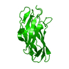
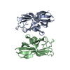
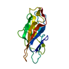
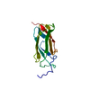
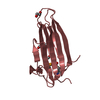
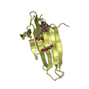
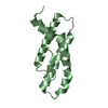
| Title | COHESIN MODULE FROM THE CELLULOSOME OF CLOSTRIDIUM CELLULOLYTICUM | ||||||
 Components Components | SCAFFOLDING PROTEIN | ||||||
 Keywords Keywords | STRUCTURAL PROTEIN / Beta -Barrel | ||||||
| Function / homology |  Function and homology information Function and homology informationcellulose binding / cellulose catabolic process / extracellular region / metal ion binding Similarity search - Function | ||||||
| Biological species |  Clostridium cellulolyticum (bacteria) Clostridium cellulolyticum (bacteria) | ||||||
| Method |  X-RAY DIFFRACTION / SYNCHROTRON / Resolution: 2 Å X-RAY DIFFRACTION / SYNCHROTRON / Resolution: 2 Å | ||||||
 Authors Authors | Spinelli, S. / Fierobe, H.-P. / Belaich, A. / Belaich, J.-P. / Henrissat, B. / Cambillau, C. | ||||||
 Citation Citation | Journal: J.Mol.Biol. / Year: 2000 Title: Crystal structure of a cohesin module from Clostridium cellulolyticum: implications for dockerin recognition. Authors: Spinelli, S. / Fierobe, H.P. / Belaich, A. / Belaich, J.P. / Henrissat, B. / Cambillau, C. | ||||||
| History |
|
- Structure visualization
| Structure viewer | Molecule: MolmilJmol/JSmol |
|---|
- Downloads & links
-Download
| PDBx/mmCIF format | 1g1k.cif.gz | 66.4 KB | Display | PDBx/mmCIF format |
|---|---|---|---|---|
| PDB format | pdb1g1k.ent.gz | 50.1 KB | Display | PDB format |
| PDBx/mmJSON format | 1g1k.json.gz | Tree view | PDBx/mmJSON format | |
| Others |  Other downloads Other downloads |
-Validation report
| Arichive directory | https://data.pdbj.org/pub/pdb/validation_reports/g1/1g1kftp://data.pdbj.org/pub/pdb/validation_reports/g1/1g1k | HTTPS FTP |
|---|
-Related structure data
| Related structure data | |
|---|---|
| Similar structure data |


-Links
 PDBj
PDBj
- Assembly
| Deposited unit | 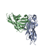
| ||||||||||
|---|---|---|---|---|---|---|---|---|---|---|---|
| 1 | 
| ||||||||||
| 2 | 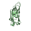
| ||||||||||
| Unit cell |
| ||||||||||
| Details | MONOMER, POSSIBLY A DIMER |
-Components
| #1: Protein | Mass: 14648.539 Da / Num. of mol.: 2 / Fragment: RESIDUES 287 - 428 Source method: isolated from a genetically manipulated source Source: (gene. exp.) Clostridium cellulolyticum (bacteria) / Tissue: CELLULOSOME MODULE / Plasmid: PET-COH 1B / Species (production host): Escherichia coli / Production host: #2: Water | ChemComp-HOH / |  Mass: 18.015 Da / Num. of mol.: 226 / Source method: isolated from a natural source / Formula: H2O Mass: 18.015 Da / Num. of mol.: 226 / Source method: isolated from a natural source / Formula: H2O |
|---|
-Experimental details
-Experiment
| Experiment | Method: X-RAY DIFFRACTION / Number of used crystals: 1 |
|---|
- Sample preparation
| Crystal | Density Matthews: 2.92 Å3/Da / Density % sol: 57.83 % | ||||||||||||||||||||
|---|---|---|---|---|---|---|---|---|---|---|---|---|---|---|---|---|---|---|---|---|---|
| Crystal grow | Temperature: 293 K / Method: vapor diffusion, hanging drop / pH: 7 Details: PEG 8000 (30%), 150 mM (NH4)2SO4, 100 mM guanidine hydrochloride, pH 7, VAPOR DIFFUSION, HANGING DROP, temperature 293K | ||||||||||||||||||||
| Crystal | *PLUS Density % sol: 50 % | ||||||||||||||||||||
| Crystal grow | *PLUS Temperature: 20 ℃ / Details: used macroseeding | ||||||||||||||||||||
| Components of the solutions | *PLUS
|
-Data collection
| Diffraction | Mean temperature: 100 K |
|---|---|
| Diffraction source | Source: SYNCHROTRON / Site: ESRF  / Beamline: ID2 / Wavelength: 0.988 / Beamline: ID2 / Wavelength: 0.988 |
| Detector | Type: MARRESEARCH / Detector: IMAGE PLATE / Date: Feb 22, 1998 |
| Radiation | Protocol: SINGLE WAVELENGTH / Monochromatic (M) / Laue (L): M / Scattering type: x-ray |
| Radiation wavelength | Wavelength: 0.988 Å / Relative weight: 1 |
| Reflection | Resolution: 2→20 Å / Num. all: 24484 / Num. obs: 22829 / % possible obs: 98 % / Observed criterion σ(F): 1 / Observed criterion σ(I): 1 / Redundancy: 3.5 % / Biso Wilson estimate: 27 Å2 / Rmerge(I) obs: 0.082 / Net I/σ(I): 6.4 |
| Reflection shell | Resolution: 2→2.06 Å / Redundancy: 3.4 % / Rmerge(I) obs: 0.275 / % possible all: 97.8 |
| Reflection | *PLUS % possible obs: 98 % |
| Reflection shell | *PLUS % possible obs: 98.8 % / Mean I/σ(I) obs: 2 |
- Processing
| Software |
| |||||||||||||||||||||||||
|---|---|---|---|---|---|---|---|---|---|---|---|---|---|---|---|---|---|---|---|---|---|---|---|---|---|---|
| Refinement | Resolution: 2→20 Å / Cross valid method: THROUGHOUT / σ(F): 0 / σ(I): 0 / Stereochemistry target values: Engh & Huber
| |||||||||||||||||||||||||
| Refinement step | Cycle: LAST / Resolution: 2→20 Å
| |||||||||||||||||||||||||
| Refine LS restraints |
| |||||||||||||||||||||||||
| Software | *PLUS Name: CNS / Classification: refinement | |||||||||||||||||||||||||
| Refinement | *PLUS Lowest resolution: 20 Å / σ(F): 0 / % reflection Rfree: 4.2 % / Rfactor obs: 0.23 / Rfactor Rfree: 0.27 / Rfactor Rwork: 0.23 | |||||||||||||||||||||||||
| Solvent computation | *PLUS | |||||||||||||||||||||||||
| Displacement parameters | *PLUS | |||||||||||||||||||||||||
| Refine LS restraints | *PLUS
|
