 Movie
Movie Controller

[English] 日本語
 Yorodumi
- PDB-1edg: SINGLE CRYSTAL STRUCTURE DETERMINATION OF THE CATALYTIC DOMAIN OF... -
+ Open data

- Basic information
| Entry | Database: PDB / ID: 1edg | ||||||
|---|---|---|---|---|---|---|---|
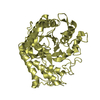
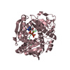
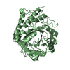
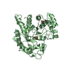
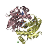
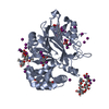
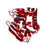
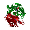
| Title | SINGLE CRYSTAL STRUCTURE DETERMINATION OF THE CATALYTIC DOMAIN OF CELCCA CARRIED OUT AT 15 DEGREE C | ||||||
 Components Components | ENDOGLUCANASE A | ||||||
 Keywords Keywords | CELLULOSE DEGRADATION / FAMILY A / CELLULASES / XYLANASES / FAMILY 5 OF GLYCOSYL HYDROLASE | ||||||
| Function / homology |  Function and homology information Function and homology informationcellulase / cellulase activity / beta-glucosidase activity / cellulose catabolic process / cell surface / extracellular region Similarity search - Function | ||||||
| Biological species |  Clostridium cellulolyticum (bacteria) Clostridium cellulolyticum (bacteria) | ||||||
| Method |  X-RAY DIFFRACTION / SYNCHROTRON / Resolution: 1.6 Å X-RAY DIFFRACTION / SYNCHROTRON / Resolution: 1.6 Å | ||||||
 Authors Authors | Ducros, V. / Czjzek, M. / Haser, R. | ||||||
 Citation Citation | Journal: Structure / Year: 1995 Title: Crystal structure of the catalytic domain of a bacterial cellulase belonging to family 5. Authors: Ducros, V. / Czjzek, M. / Belaich, A. / Gaudin, C. / Fierobe, H.P. / Belaich, J.P. / Davies, G.J. / Haser, R. #1: Journal: J.Bacteriol. / Year: 1991Title: Characterization of Endoglucanase a from Clostridium Cellulolyticum Authors: Fierobe, H.P. / Gaudin, C. / Belaich, A. / Loutfi, M. / Faure, E. / Bagnara, C. / Baty, D. / Belaich, J.P. | ||||||
| History |
|
- Structure visualization
| Structure viewer | Molecule: MolmilJmol/JSmol |
|---|
- Downloads & links
-Download
| PDBx/mmCIF format | 1edg.cif.gz | 94.2 KB | Display | PDBx/mmCIF format |
|---|---|---|---|---|
| PDB format | pdb1edg.ent.gz | 71.6 KB | Display | PDB format |
| PDBx/mmJSON format | 1edg.json.gz | Tree view | PDBx/mmJSON format | |
| Others |  Other downloads Other downloads |
-Validation report
| Arichive directory | https://data.pdbj.org/pub/pdb/validation_reports/ed/1edgftp://data.pdbj.org/pub/pdb/validation_reports/ed/1edg | HTTPS FTP |
|---|
-Related structure data
| Similar structure data |
|---|


-Links
 PDBj
PDBj- Assembly
| Deposited unit | 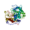
| ||||||||
|---|---|---|---|---|---|---|---|---|---|
| 1 |
| ||||||||
| Unit cell |
|
-Components
| #1: Protein | Mass: 43135.414 Da / Num. of mol.: 1 / Fragment: CATALYTIC DOMAIN, C TERMINUS TRUNCATED Source method: isolated from a genetically manipulated source Details: FAMILY 5 OF GLYCOSYL HYDROLASES / Source: (gene. exp.) Clostridium cellulolyticum (bacteria) / Strain: H10 / Gene: CELCCA / Plasmid: PJF118EH / Gene (production host): CELCCA / Production host: |
|---|---|
| #2: Water | ChemComp-HOH /  Mass: 18.015 Da / Num. of mol.: 375 / Source method: isolated from a natural source / Formula: H2O Mass: 18.015 Da / Num. of mol.: 375 / Source method: isolated from a natural source / Formula: H2O |
-Experimental details
-Experiment
| Experiment | Method: X-RAY DIFFRACTION |
|---|
- Sample preparation
| Crystal | Density Matthews: 2.64 Å3/Da / Density % sol: 53 % | ||||||||||||||||||||||||||||||
|---|---|---|---|---|---|---|---|---|---|---|---|---|---|---|---|---|---|---|---|---|---|---|---|---|---|---|---|---|---|---|---|
| Crystal grow | Temperature: 277 K / pH: 6 Details: CRYSTALS WERE GROWN AT 4 DEGREES C AND PH 6.0, temperature 277K | ||||||||||||||||||||||||||||||
| Crystal | *PLUS | ||||||||||||||||||||||||||||||
| Crystal grow | *PLUS Temperature: 4 ℃ / Method: vapor diffusion, hanging drop / Details: Roig, V., (1993) J. Mol. Biol., 233, 325. | ||||||||||||||||||||||||||||||
| Components of the solutions | *PLUS
|
-Data collection
| Diffraction |
| |||||||||||||||
|---|---|---|---|---|---|---|---|---|---|---|---|---|---|---|---|---|
| Diffraction source |
| |||||||||||||||
| Detector |
| |||||||||||||||
| Radiation |
| |||||||||||||||
| Radiation wavelength |
| |||||||||||||||
| Reflection | Resolution: 1.6→20 Å / Num. obs: 59967 / % possible obs: 98 % / Observed criterion σ(I): 2.5 / Redundancy: 5.9 % / Rmerge(I) obs: 0.069 | |||||||||||||||
| Reflection | *PLUS Highest resolution: 1.6 Å / Num. measured all: 355167 / Rmerge(I) obs: 0.072 |

- Processing
| Software |
| ||||||||||||||||||||||||||||||||||||||||||||||||||||||||||||
|---|---|---|---|---|---|---|---|---|---|---|---|---|---|---|---|---|---|---|---|---|---|---|---|---|---|---|---|---|---|---|---|---|---|---|---|---|---|---|---|---|---|---|---|---|---|---|---|---|---|---|---|---|---|---|---|---|---|---|---|---|---|
| Refinement | Resolution: 1.6→7 Å / σ(F): 2 Details: THE SIDE CHAIN ATOMS OF TRP 181 AND ARG 358 WERE MODELED WITH PARTIAL OCCUPATION OF 0.7. THE SIDE CHAIN ATOMS OF ASN 46 WERE MODELED WITH PARTIAL OCCUPATION OF 0.6.
| ||||||||||||||||||||||||||||||||||||||||||||||||||||||||||||
| Displacement parameters | Biso mean: 17.35 Å2 | ||||||||||||||||||||||||||||||||||||||||||||||||||||||||||||
| Refine analyze | Luzzati coordinate error obs: 0.17 Å | ||||||||||||||||||||||||||||||||||||||||||||||||||||||||||||
| Refinement step | Cycle: LAST / Resolution: 1.6→7 Å
| ||||||||||||||||||||||||||||||||||||||||||||||||||||||||||||
| Refine LS restraints |
| ||||||||||||||||||||||||||||||||||||||||||||||||||||||||||||
| Software | *PLUS Name: X-PLOR / Version: 3.1 / Classification: refinement | ||||||||||||||||||||||||||||||||||||||||||||||||||||||||||||
| Refinement | *PLUS Rfactor Rfree: 0.2315 | ||||||||||||||||||||||||||||||||||||||||||||||||||||||||||||
| Solvent computation | *PLUS | ||||||||||||||||||||||||||||||||||||||||||||||||||||||||||||
| Displacement parameters | *PLUS | ||||||||||||||||||||||||||||||||||||||||||||||||||||||||||||
| Refine LS restraints | *PLUS
|
