 Movie
Movie Controller


+ Open data

- Basic information
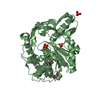
| Entry | Database: PDB / ID: 2jep | ||||||
|---|---|---|---|---|---|---|---|
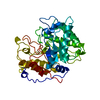
| Title | Native family 5 xyloglucanase from Paenibacillus pabuli | ||||||
 Components Components | XYLOGLUCANASE | ||||||
 Keywords Keywords | HYDROLASE / FAMILY 5 / XYLOGLUCANASE / PLANT CELL WALL | ||||||
| Function / homology |  Function and homology information Function and homology informationxyloglucan-specific endo-beta-1,4-glucanase / xyloglucan-specific endo-beta-1,4-glucanase activity / beta-glucosidase activity / cellulose catabolic process / cell surface / extracellular region Similarity search - Function | ||||||
| Biological species |  PAENIBACILLUS PABULI (bacteria) PAENIBACILLUS PABULI (bacteria) | ||||||
| Method |  X-RAY DIFFRACTION / SYNCHROTRON / MOLECULAR REPLACEMENT / Resolution: 1.4 Å X-RAY DIFFRACTION / SYNCHROTRON / MOLECULAR REPLACEMENT / Resolution: 1.4 Å | ||||||
 Authors Authors | Gloster, T.M. / Ibatullin, F.M. / Macauley, K. / Eklof, J.M. / Roberts, S. / Turkenburg, J.P. / Bjornvad, M.E. / Jorgensen, P.L. / Danielsen, S. / Johansen, K. ...Gloster, T.M. / Ibatullin, F.M. / Macauley, K. / Eklof, J.M. / Roberts, S. / Turkenburg, J.P. / Bjornvad, M.E. / Jorgensen, P.L. / Danielsen, S. / Johansen, K. / Borchert, T.V. / Wilson, K.S. / Brumer, H. / Davies, G.J. | ||||||
 Citation Citation | Journal: J.Biol.Chem. / Year: 2007 Title: Characterization and Three-Dimensional Structures of Two Distinct Bacterial Xyloglucanases from Families Gh5 and Gh12. Authors: Gloster, T.M. / Ibatullin, F.M. / Macauley, K. / Eklof, J.M. / Roberts, S. / Turkenburg, J.P. / Bjornvad, M.E. / Jorgensen, P.L. / Danielsen, S. / Johansen, K. / Borchert, T.V. / Wilson, K.S. ...Authors: Gloster, T.M. / Ibatullin, F.M. / Macauley, K. / Eklof, J.M. / Roberts, S. / Turkenburg, J.P. / Bjornvad, M.E. / Jorgensen, P.L. / Danielsen, S. / Johansen, K. / Borchert, T.V. / Wilson, K.S. / Brumer, H. / Davies, G.J. | ||||||
| History |
| ||||||
| Remark 700 | SHEET DETERMINATION METHOD: DSSP THE SHEETS PRESENTED AS "AA" IN EACH CHAIN ON SHEET RECORDS BELOW ... SHEET DETERMINATION METHOD: DSSP THE SHEETS PRESENTED AS "AA" IN EACH CHAIN ON SHEET RECORDS BELOW IS ACTUALLY AN 9-STRANDED BARREL THIS IS REPRESENTED BY A 10-STRANDED SHEET IN WHICH THE FIRST AND LAST STRANDS ARE IDENTICAL. THE SHEETS PRESENTED AS "BA" IN EACH CHAIN ON SHEET RECORDS BELOW IS ACTUALLY AN 8-STRANDED BARREL THIS IS REPRESENTED BY A 9-STRANDED SHEET IN WHICH THE FIRST AND LAST STRANDS ARE IDENTICAL. |
- Structure visualization
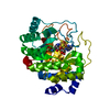
| Structure viewer | Molecule: MolmilJmol/JSmol |
|---|
- Downloads & links
-Download
| PDBx/mmCIF format | 2jep.cif.gz | 349.8 KB | Display | PDBx/mmCIF format |
|---|---|---|---|---|
| PDB format | pdb2jep.ent.gz | 286 KB | Display | PDB format |
| PDBx/mmJSON format | 2jep.json.gz | Tree view | PDBx/mmJSON format | |
| Others |  Other downloads Other downloads |
-Validation report
| Arichive directory | https://data.pdbj.org/pub/pdb/validation_reports/je/2jepftp://data.pdbj.org/pub/pdb/validation_reports/je/2jep | HTTPS FTP |
|---|
-Related structure data
| Related structure data | 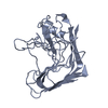 2jemC 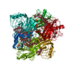 2jenC  2jeqC  1ecgS S: Starting model for refinement C: citing same article ( |
|---|---|
| Similar structure data |


-Links
 PDBj
PDBj- Assembly
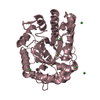
| Deposited unit | 
| ||||||||
|---|---|---|---|---|---|---|---|---|---|
| 1 | 
| ||||||||
| 2 | 
| ||||||||
| Unit cell |
| ||||||||
| Noncrystallographic symmetry (NCS) | NCS oper: (Code: given Matrix: (0.9999, -0.01231, 0.009834), Vector: |
-Components
| #1: Protein | Mass: 43969.871 Da / Num. of mol.: 2 Source method: isolated from a genetically manipulated source Source: (gene. exp.) PAENIBACILLUS PABULI (bacteria) / Strain: DSM13330 / Production host: References: UniProt: H9KVH3*PLUS, xyloglucan-specific endo-beta-1,4-glucanase #2: Chemical |   Mass: 62.068 Da / Num. of mol.: 3 / Source method: obtained synthetically / Formula: C2H6O2 Mass: 62.068 Da / Num. of mol.: 3 / Source method: obtained synthetically / Formula: C2H6O2#3: Chemical |   Mass: 40.078 Da / Num. of mol.: 2 / Source method: obtained synthetically / Formula: Ca Mass: 40.078 Da / Num. of mol.: 2 / Source method: obtained synthetically / Formula: Ca#4: Water | ChemComp-HOH / |  Mass: 18.015 Da / Num. of mol.: 1278 / Source method: isolated from a natural source / Formula: H2O Mass: 18.015 Da / Num. of mol.: 1278 / Source method: isolated from a natural source / Formula: H2OSequence details | THE FIRST 32 RESIDUES CANNOT BE OBSERVED, AND ARE LIKELY TO CORRESPOND TO A SIGNAL PEPTIDE CLEAVED ...THE FIRST 32 RESIDUES CANNOT BE OBSERVED, AND ARE LIKELY TO CORRESPOND | |
|---|
-Experimental details
-Experiment
| Experiment | Method: X-RAY DIFFRACTION / Number of used crystals: 1 |
|---|
- Sample preparation
| Crystal | Density Matthews: 2.1 Å3/Da / Density % sol: 41.7 % |
|---|---|
| Crystal grow | pH: 8.5 / Details: 20-25% PEG 8000, 0.2 M CACL2, 0.1 M TRIS, PH 8.5 |
-Data collection
| Diffraction | Mean temperature: 100 K |
|---|---|
| Diffraction source | Source: SYNCHROTRON / Site: ESRF  / Beamline: ID14-3 / Wavelength: 0.931 / Beamline: ID14-3 / Wavelength: 0.931 |
| Detector | Type: ADSC CCD / Detector: CCD / Details: TOROIDAL MIRROR |
| Radiation | Monochromator: DIAMOND (111), GE(220) / Protocol: SINGLE WAVELENGTH / Monochromatic (M) / Laue (L): M / Scattering type: x-ray |
| Radiation wavelength | Wavelength: 0.931 Å / Relative weight: 1 |
| Reflection | Resolution: 1.4→30 Å / Num. obs: 134851 / % possible obs: 99.7 % / Redundancy: 4.4 % / Rmerge(I) obs: 0.04 / Net I/σ(I): 42.2 |
| Reflection shell | Resolution: 1.4→1.42 Å / Redundancy: 3.2 % / Rmerge(I) obs: 0.11 / Mean I/σ(I) obs: 9.1 / % possible all: 98.6 |
- Processing
| Software |
| ||||||||||||||||||||||||||||||||||||||||||||||||||||||||||||||||||||||||||||||||||||||||||||||||||||||||||||||||||||||||||||||||||||||||||||||||||||||||||||||||||||||||||||||||||||||
|---|---|---|---|---|---|---|---|---|---|---|---|---|---|---|---|---|---|---|---|---|---|---|---|---|---|---|---|---|---|---|---|---|---|---|---|---|---|---|---|---|---|---|---|---|---|---|---|---|---|---|---|---|---|---|---|---|---|---|---|---|---|---|---|---|---|---|---|---|---|---|---|---|---|---|---|---|---|---|---|---|---|---|---|---|---|---|---|---|---|---|---|---|---|---|---|---|---|---|---|---|---|---|---|---|---|---|---|---|---|---|---|---|---|---|---|---|---|---|---|---|---|---|---|---|---|---|---|---|---|---|---|---|---|---|---|---|---|---|---|---|---|---|---|---|---|---|---|---|---|---|---|---|---|---|---|---|---|---|---|---|---|---|---|---|---|---|---|---|---|---|---|---|---|---|---|---|---|---|---|---|---|---|---|
| Refinement | Method to determine structure: MOLECULAR REPLACEMENT Starting model: PDB ENTRY 1ECG Resolution: 1.4→63.25 Å / Cor.coef. Fo:Fc: 0.982 / Cor.coef. Fo:Fc free: 0.976 / SU B: 1.272 / SU ML: 0.024 / Cross valid method: THROUGHOUT / ESU R: 0.056 / ESU R Free: 0.049 / Stereochemistry target values: MAXIMUM LIKELIHOOD Details: HYDROGENS HAVE BEEN ADDED IN THE RIDING POSITIONS. RESIDUES 34-37 IN THE B CHAIN DO NOT FIT THE DENSITY WELL, BUT HAVE BEEN MODELLED AS IN THE A CHAIN STRUCTURE WHICH IS MORE ORDERED. THERE ...Details: HYDROGENS HAVE BEEN ADDED IN THE RIDING POSITIONS. RESIDUES 34-37 IN THE B CHAIN DO NOT FIT THE DENSITY WELL, BUT HAVE BEEN MODELLED AS IN THE A CHAIN STRUCTURE WHICH IS MORE ORDERED. THERE IS ALSO DENSITY IN THE ACTIVE SITE (NEAR TO RESIDUES 182 & 323) WHICH CANNOT BE MODELLED AS ANY SMALL MOLECULE KNOWN TO BE PRESENT AND HAS INSTEAD CONTAINS WATER MOLECULES.
| ||||||||||||||||||||||||||||||||||||||||||||||||||||||||||||||||||||||||||||||||||||||||||||||||||||||||||||||||||||||||||||||||||||||||||||||||||||||||||||||||||||||||||||||||||||||
| Solvent computation | Ion probe radii: 0.8 Å / Shrinkage radii: 0.8 Å / VDW probe radii: 1.4 Å / Solvent model: MASK | ||||||||||||||||||||||||||||||||||||||||||||||||||||||||||||||||||||||||||||||||||||||||||||||||||||||||||||||||||||||||||||||||||||||||||||||||||||||||||||||||||||||||||||||||||||||
| Displacement parameters | Biso mean: 8.77 Å2
| ||||||||||||||||||||||||||||||||||||||||||||||||||||||||||||||||||||||||||||||||||||||||||||||||||||||||||||||||||||||||||||||||||||||||||||||||||||||||||||||||||||||||||||||||||||||
| Refinement step | Cycle: LAST / Resolution: 1.4→63.25 Å
| ||||||||||||||||||||||||||||||||||||||||||||||||||||||||||||||||||||||||||||||||||||||||||||||||||||||||||||||||||||||||||||||||||||||||||||||||||||||||||||||||||||||||||||||||||||||
| Refine LS restraints |
|