登録情報 データベース : PDB / ID : 2jenタイトル Family 12 xyloglucanase from Bacillus licheniformis in complex with ligand ENDO-BETA-1,4-GLUCANASE キーワード / / / / 機能・相同性 分子機能 ドメイン・相同性 構成要素
/ / / / / / / / / 生物種 BACILLUS LICHENIFORMIS (バクテリア)手法 / / / 解像度 : 1.4 Å データ登録者 Gloster, T.M. / Ibatullin, F.M. / Macauley, K. / Eklof, J.M. / Roberts, S. / Turkenburg, J.P. / Bjornvad, M.E. / Jorgensen, P.L. / Danielsen, S. / Johansen, K.S. ...Gloster, T.M. / Ibatullin, F.M. / Macauley, K. / Eklof, J.M. / Roberts, S. / Turkenburg, J.P. / Bjornvad, M.E. / Jorgensen, P.L. / Danielsen, S. / Johansen, K.S. / Borchert, T.V. / Wilson, K.S. / Brumer, H. / Davies, G.J. ジャーナル : J.Biol.Chem. / 年 : 2007タイトル : Characterization and Three-Dimensional Structures of Two Distinct Bacterial Xyloglucanases from Families Gh5 and Gh12.著者: Gloster, T.M. / Ibatullin, F.M. / Macauley, K. / Eklof, J.M. / Roberts, S. / Turkenburg, J.P. / Bjornvad, M.E. / Jorgensen, P.L. / Danielsen, S. / Johansen, K.S. / Borchert, T.V. / Wilson, K. ... 著者 : Gloster, T.M. / Ibatullin, F.M. / Macauley, K. / Eklof, J.M. / Roberts, S. / Turkenburg, J.P. / Bjornvad, M.E. / Jorgensen, P.L. / Danielsen, S. / Johansen, K.S. / Borchert, T.V. / Wilson, K.S. / Brumer, H. / Davies, G.J. 履歴 登録 2007年1月18日 登録サイト / 処理サイト 改定 1.0 2007年3月20日 Provider / タイプ 改定 1.1 2011年5月8日 Group 改定 1.2 2011年7月13日 Group 改定 2.0 2020年7月29日 Group Advisory / Atomic model ... Advisory / Atomic model / Data collection / Derived calculations / Other / Structure summary カテゴリ atom_site / atom_site_anisotrop ... atom_site / atom_site_anisotrop / chem_comp / database_PDB_caveat / entity / pdbx_branch_scheme / pdbx_chem_comp_identifier / pdbx_database_status / pdbx_entity_branch / pdbx_entity_branch_descriptor / pdbx_entity_branch_link / pdbx_entity_branch_list / pdbx_entity_nonpoly / pdbx_nonpoly_scheme / pdbx_struct_assembly_gen / pdbx_validate_chiral / struct_asym / struct_conn / struct_site / struct_site_gen Item _atom_site.B_iso_or_equiv / _atom_site.Cartn_x ... _atom_site.B_iso_or_equiv / _atom_site.Cartn_x / _atom_site.Cartn_y / _atom_site.Cartn_z / _atom_site.auth_asym_id / _atom_site.auth_atom_id / _atom_site.auth_comp_id / _atom_site.auth_seq_id / _atom_site.label_alt_id / _atom_site.label_asym_id / _atom_site.label_atom_id / _atom_site.label_comp_id / _atom_site.label_entity_id / _atom_site.occupancy / _atom_site.type_symbol / _atom_site_anisotrop.U[1][1] / _atom_site_anisotrop.U[1][2] / _atom_site_anisotrop.U[1][3] / _atom_site_anisotrop.U[2][2] / _atom_site_anisotrop.U[2][3] / _atom_site_anisotrop.U[3][3] / _atom_site_anisotrop.pdbx_auth_asym_id / _atom_site_anisotrop.pdbx_auth_atom_id / _atom_site_anisotrop.pdbx_auth_comp_id / _atom_site_anisotrop.pdbx_auth_seq_id / _atom_site_anisotrop.pdbx_label_alt_id / _atom_site_anisotrop.pdbx_label_asym_id / _atom_site_anisotrop.pdbx_label_atom_id / _atom_site_anisotrop.pdbx_label_comp_id / _atom_site_anisotrop.type_symbol / _chem_comp.name / _chem_comp.type / _pdbx_database_status.status_code_sf / _pdbx_struct_assembly_gen.asym_id_list / _pdbx_validate_chiral.auth_asym_id / _pdbx_validate_chiral.auth_seq_id / _struct_conn.pdbx_dist_value / _struct_conn.pdbx_leaving_atom_flag / _struct_conn.ptnr1_auth_asym_id / _struct_conn.ptnr1_auth_seq_id / _struct_conn.ptnr1_label_asym_id / _struct_conn.ptnr1_label_atom_id / _struct_conn.ptnr2_auth_asym_id / _struct_conn.ptnr2_auth_comp_id / _struct_conn.ptnr2_auth_seq_id / _struct_conn.ptnr2_label_asym_id / _struct_conn.ptnr2_label_comp_id 解説 / Provider / タイプ 改定 2.1 2023年12月13日 Group Data collection / Database references ... Data collection / Database references / Refinement description / Structure summary カテゴリ chem_comp / chem_comp_atom ... chem_comp / chem_comp_atom / chem_comp_bond / database_2 / pdbx_initial_refinement_model Item / _database_2.pdbx_DOI / _database_2.pdbx_database_accession
すべて表示 表示を減らす Remark 700 SHEET THE SHEET STRUCTURE OF THIS MOLECULE IS BIFURCATED. IN ORDER TO REPRESENT THIS FEATURE IN ... SHEET THE SHEET STRUCTURE OF THIS MOLECULE IS BIFURCATED. IN ORDER TO REPRESENT THIS FEATURE IN THE SHEET RECORDS BELOW, TWO SHEETS ARE DEFINED.
 ムービー
ムービー コントローラー


 データを開く
 基本情報
基本情報 要素
要素 キーワード
キーワード 機能・相同性情報
 X線回折 /
  データ登録者
 引用
引用 構造の表示
構造の表示 ダウンロードとリンク
 その他のダウンロード
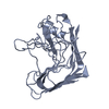


 PDBj
PDBj
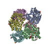
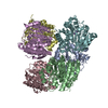
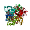
 集合体
集合体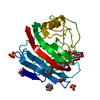


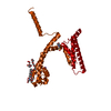
 分子量: 88.105 Da / 分子数: 2 / 由来タイプ: 合成 / 式: C4H8O2
 分子量: 92.094 Da / 分子数: 1 / 由来タイプ: 合成 / 式: C3H8O3
 分子量: 96.063 Da / 分子数: 2 / 由来タイプ: 合成 / 式: SO4
 試料調製
試料調製 / ビームライン: ID14-1 / 波長: 0.934
  解析
解析