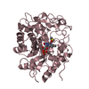
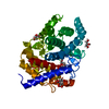
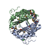
methylornithine synthase / pyrrolysine biosynthetic process / intramolecular transferase activity / transferase activity / 4 iron, 4 sulfur cluster binding / metal ion binding Similarity search - Function
Pyrrolysine biosynthesis protein PylB / HydE/PylB-like / Elp3/MiaB/NifB / Elongator protein 3, MiaB family, Radical SAM / : / Radical SAM superfamily / Radical SAM core domain profile. / Radical SAM / Aldolase class I / Aldolase-type TIM barrel ...Pyrrolysine biosynthesis protein PylB / HydE/PylB-like / Elp3/MiaB/NifB / Elongator protein 3, MiaB family, Radical SAM / : / Radical SAM superfamily / Radical SAM core domain profile. / Radical SAM / Aldolase class I / Aldolase-type TIM barrel / TIM Barrel / Alpha-Beta Barrel / Alpha Beta Similarity search - Domain/homology
Resolution: 1.5→1.6 Å / Redundancy: 5.3 % / Rmerge(I) obs: 0.539 / Mean I/σ(I) obs: 3.2 / Rsym value: 0.525 / % possible all: 99.9
-
Processing
Software
Name
Classification
XDS
datascaling
REFMAC
refinement
XDS
datareduction
XSCALE
datascaling
REFMAC
phasing
Refinement
Method to determine structure: MOLECULAR REPLACEMENT / Resolution: 1.5→10 Å / Cor.coef. Fo:Fc: 0.976 / Cor.coef. Fo:Fc free: 0.966 / SU B: 1.977 / SU ML: 0.034 / Cross valid method: THROUGHOUT / ESU R: 0.065 / ESU R Free: 0.059 / Stereochemistry target values: MAXIMUM LIKELIHOOD / Details: HYDROGENS HAVE BEEN USED IF PRESENT IN THE INPUT
Rfactor
Num. reflection
% reflection
Selection details
Rfree
0.16504
2904
5 %
RANDOM
Rwork
0.12865
-
-
-
obs
0.13049
55170
99.83 %
-
all
-
57839
-
-
Solvent computation
Ion probe radii: 0.8 Å / Shrinkage radii: 0.8 Å / VDW probe radii: 1.2 Å / Solvent model: MASK
Displacement parameters
Biso mean: 25.247 Å2
Baniso -1
Baniso -2
Baniso -3
1-
0.01 Å2
0.01 Å2
0 Å2
2-
-
0.01 Å2
0 Å2
3-
-
-
-0.02 Å2
Refinement step
Cycle: LAST / Resolution: 1.5→10 Å
Protein
Nucleic acid
Ligand
Solvent
Total
Num. atoms
2699
0
45
315
3059
Refine LS restraints
Refine-ID
Type
Dev ideal
Dev ideal target
Number
X-RAY DIFFRACTION
r_bond_refined_d
0.029
0.019
2804
X-RAY DIFFRACTION
r_bond_other_d
X-RAY DIFFRACTION
r_angle_refined_deg
2.576
2.016
3828
X-RAY DIFFRACTION
r_angle_other_deg
X-RAY DIFFRACTION
r_dihedral_angle_1_deg
5.96
5
336
X-RAY DIFFRACTION
r_dihedral_angle_2_deg
35.165
23.676
136
X-RAY DIFFRACTION
r_dihedral_angle_3_deg
12.872
15
510
X-RAY DIFFRACTION
r_dihedral_angle_4_deg
15.308
15
27
X-RAY DIFFRACTION
r_chiral_restr
0.289
0.2
420
X-RAY DIFFRACTION
r_gen_planes_refined
0.013
0.021
2096
X-RAY DIFFRACTION
r_gen_planes_other
X-RAY DIFFRACTION
r_nbd_refined
X-RAY DIFFRACTION
r_nbd_other
X-RAY DIFFRACTION
r_nbtor_refined
X-RAY DIFFRACTION
r_nbtor_other
X-RAY DIFFRACTION
r_xyhbond_nbd_refined
X-RAY DIFFRACTION
r_xyhbond_nbd_other
X-RAY DIFFRACTION
r_metal_ion_refined
X-RAY DIFFRACTION
r_metal_ion_other
X-RAY DIFFRACTION
r_symmetry_vdw_refined
X-RAY DIFFRACTION
r_symmetry_vdw_other
X-RAY DIFFRACTION
r_symmetry_hbond_refined
X-RAY DIFFRACTION
r_symmetry_hbond_other
X-RAY DIFFRACTION
r_symmetry_metal_ion_refined
X-RAY DIFFRACTION
r_symmetry_metal_ion_other
X-RAY DIFFRACTION
r_mcbond_it
1.418
1.5
1683
X-RAY DIFFRACTION
r_mcbond_other
X-RAY DIFFRACTION
r_mcangle_it
2.529
2
2713
X-RAY DIFFRACTION
r_scbond_it
3.188
3
1115
X-RAY DIFFRACTION
r_scangle_it
4.963
4.5
1055
X-RAY DIFFRACTION
r_rigid_bond_restr
8.312
3
2802
X-RAY DIFFRACTION
r_sphericity_free
36.259
5
101
X-RAY DIFFRACTION
r_sphericity_bonded
14.847
5
2963
LS refinement shell
Resolution: 1.5→1.538 Å / Total num. of bins used: 20
Rfactor
Num. reflection
% reflection
Rfree
0.221
199
-
Rwork
0.161
3718
-
obs
-
-
99.77 %
Refinement TLS params.
Method: refined / Refine-ID: X-RAY DIFFRACTION
ID
L11 (°2)
L12 (°2)
L13 (°2)
L22 (°2)
L23 (°2)
L33 (°2)
S11 (Å °)
S12 (Å °)
S13 (Å °)
S21 (Å °)
S22 (Å °)
S23 (Å °)
S31 (Å °)
S32 (Å °)
S33 (Å °)
T11 (Å2)
T12 (Å2)
T13 (Å2)
T22 (Å2)
T23 (Å2)
T33 (Å2)
Origin x (Å)
Origin y (Å)
Origin z (Å)
1
2.1219
1.4642
-1.9274
1.0135
-1.3329
1.7544
0.3563
-0.3783
-0.0018
0.2763
-0.3039
0.0149
-0.3578
0.3388
-0.0524
0.1772
-0.1108
0.0424
0.1565
-0.0176
0.0916
44.2078
45.9911
28.9195
2
0.2413
0.1358
-0.2493
0.3478
0.0998
0.4797
0.0454
0.0453
0.0781
0.0105
0.0326
0.0632
-0.046
-0.0186
-0.078
0.1022
0.0061
0.0194
0.0972
0.0087
0.102
46.25
45.5562
9.4658
3
0.2894
0.1652
-0.134
0.2517
-0.0352
0.141
-0.0193
0.0281
-0.0202
0.0213
0.0291
-0.0326
0.001
-0.0075
-0.0098
0.1104
-0.0135
-0.0001
0.108
-0.0053
0.0936
57.827
33.5713
-1.0695
4
0.316
0.8319
-0.3168
2.2931
-0.8818
0.3403
0.0243
-0.0515
-0.0659
0.1032
-0.0557
-0.1013
-0.0436
0.0066
0.0315
0.1211
-0.0006
-0.0155
0.1454
0.0259
0.1075
55.8764
27.1283
12.5373
5
0.7979
0.03
-0.2879
0.0567
-0.0491
0.148
0.01
-0.0104
0.0183
0.0615
0.0028
0.0015
-0.014
-0.0016
-0.0128
0.1247
-0.0067
0.0024
0.1179
-0.009
0.1001
49.6308
34.4005
15.3123
6
0.3317
0.1046
0.3705
0.0848
0.2767
1.0723
-0.026
0.0297
0.09
0.0216
-0.0059
0.0257
-0.0233
-0.0843
0.0319
0.0965
-0.0103
0.0044
0.131
0.0046
0.0976
40.859
37.0055
2.2791
7
0.437
0.2351
-0.3016
0.4133
0.034
0.3436
0.1033
0.0393
0.1311
0.087
-0.0113
0.0809
-0.0461
-0.0462
-0.0921
0.1122
0.007
0.0254
0.1015
-0.0025
0.1081
42.8931
46.0338
10.519
8
0.386
0.3218
-0.1349
0.64
0.0226
0.0974
0.0377
0.0694
0.0879
0.0136
0.0142
0.0516
-0.0208
-0.04
-0.0519
0.1087
0.0101
0.0091
0.0961
0.023
0.113
51.7392
50.0544
-0.5622
Refinement TLS group
ID
Refine-ID
Refine TLS-ID
Auth asym-ID
Auth seq-ID
1
X-RAY DIFFRACTION
1
A
12 - 29
2
X-RAY DIFFRACTION
2
A
30 - 80
3
X-RAY DIFFRACTION
3
A
81 - 188
4
X-RAY DIFFRACTION
4
A
189 - 195
5
X-RAY DIFFRACTION
5
A
196 - 232
6
X-RAY DIFFRACTION
6
A
233 - 247
7
X-RAY DIFFRACTION
7
A
248 - 295
8
X-RAY DIFFRACTION
8
A
296 - 348
9
X-RAY DIFFRACTION
8
A
991 - 993
+
About Yorodumi
-
News
-
Feb 9, 2022. New format data for meta-information of EMDB entries
New format data for meta-information of EMDB entries
Version 3 of the EMDB header file is now the official format.
The previous official version 1.9 will be removed from the archive.
In the structure databanks used in Yorodumi, some data are registered as the other names, "COVID-19 virus" and "2019-nCoV". Here are the details of the virus and the list of structure data.
Jan 31, 2019. EMDB accession codes are about to change! (news from PDBe EMDB page)
EMDB accession codes are about to change! (news from PDBe EMDB page)
The allocation of 4 digits for EMDB accession codes will soon come to an end. Whilst these codes will remain in use, new EMDB accession codes will include an additional digit and will expand incrementally as the available range of codes is exhausted. The current 4-digit format prefixed with “EMD-” (i.e. EMD-XXXX) will advance to a 5-digit format (i.e. EMD-XXXXX), and so on. It is currently estimated that the 4-digit codes will be depleted around Spring 2019, at which point the 5-digit format will come into force.
The EM Navigator/Yorodumi systems omit the EMD- prefix.
Related info.:Q: What is EMD? / ID/Accession-code notation in Yorodumi/EM Navigator
Yorodumi is a browser for structure data from EMDB, PDB, SASBDB, etc.
This page is also the successor to EM Navigator detail page, and also detail information page/front-end page for Omokage search.
The word "yorodu" (or yorozu) is an old Japanese word meaning "ten thousand". "mi" (miru) is to see.
Related info.:EMDB / PDB / SASBDB / Comparison of 3 databanks / Yorodumi Search / Aug 31, 2016. New EM Navigator & Yorodumi / Yorodumi Papers / Jmol/JSmol / Function and homology information / Changes in new EM Navigator and Yorodumi
 Movie
Movie Controller


 Open data
 Basic information
 Components
 Keywords
 Function and homology information
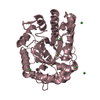
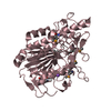
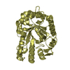
 Methanosarcina barkeri (archaea)
 X-RAY DIFFRACTION /
  Authors
 Citation
 Structure visualization
 Downloads & links
 Other downloads


 PDBj
PDBj
 Assembly
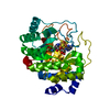


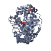
 Mass: 351.640 Da / Num. of mol.: 1 / Source method: obtained synthetically / Formula: Fe4S4
 Mass: 398.437 Da / Num. of mol.: 1 / Source method: obtained synthetically / Formula: C15H22N6O5S
 Type: D-peptide linking / Mass: 146.188 Da / Num. of mol.: 1 / Source method: obtained synthetically / Formula: C6H14N2O2
 Mass: 18.015 Da / Num. of mol.: 315 / Source method: isolated from a natural source / Formula: H2O
 Sample preparation
 / Beamline: X06DA / Wavelength: 1 Å
 Processing