 Movie
Movie Controller


+ Open data

- Basic information
| Entry | Database: PDB / ID: 6z69 | ||||||
|---|---|---|---|---|---|---|---|
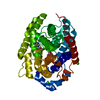
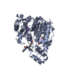
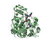
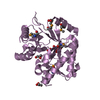
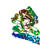
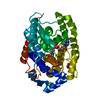
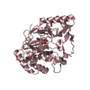
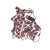
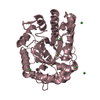
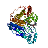
| Title | A novel metagenomic alpha/beta-fold esterase | ||||||
 Components Components | Acetyl esterase/lipase | ||||||
 Keywords Keywords | HYDROLASE / alpha/beta-fold hydrolase / esterase / metagenome / inhibitor-bound / substrate promiscuity | ||||||
| Function / homology | : / Alpha/beta hydrolase fold-3 / alpha/beta hydrolase fold / Alpha/Beta hydrolase fold / hydrolase activity / 7-hydroxy-4-methyl-2H-chromen-2-one / N-HEXYLPHOSPHONATE ETHYL ESTER / Acetyl esterase/lipase Function and homology information Function and homology information | ||||||
| Biological species |  Pseudonocardia thermophila (bacteria) Pseudonocardia thermophila (bacteria) | ||||||
| Method |  X-RAY DIFFRACTION / SYNCHROTRON / MOLECULAR REPLACEMENT / Resolution: 1.81 Å X-RAY DIFFRACTION / SYNCHROTRON / MOLECULAR REPLACEMENT / Resolution: 1.81 Å | ||||||
 Authors Authors | Bollinger, A. / Thies, S. / Hoeppner, A. / Kobus, S. / Jaeger, K.-E. / Smits, S.H.J. | ||||||
 Citation Citation | Journal: Febs J. / Year: 2021 Title: Crystal structures of a novel family IV esterase in free and substrate-bound form. Authors: Hoppner, A. / Bollinger, A. / Kobus, S. / Thies, S. / Coscolin, C. / Ferrer, M. / Jaeger, K.E. / Smits, S.H.J. | ||||||
| History |
|
- Structure visualization
| Structure viewer | Molecule: MolmilJmol/JSmol |
|---|
- Downloads & links
-Download
| PDBx/mmCIF format | 6z69.cif.gz | 174 KB | Display | PDBx/mmCIF format |
|---|---|---|---|---|
| PDB format | pdb6z69.ent.gz | 132.8 KB | Display | PDB format |
| PDBx/mmJSON format | 6z69.json.gz | Tree view | PDBx/mmJSON format | |
| Others |  Other downloads Other downloads |
-Validation report
| Arichive directory | https://data.pdbj.org/pub/pdb/validation_reports/z6/6z69ftp://data.pdbj.org/pub/pdb/validation_reports/z6/6z69 | HTTPS FTP |
|---|
-Related structure data
| Related structure data | 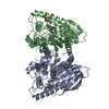 6z68SC S: Starting model for refinement C: citing same article ( |
|---|---|
| Similar structure data |
-Links
 PDBj
PDBj
- Assembly
| Deposited unit | 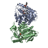
| ||||||||
|---|---|---|---|---|---|---|---|---|---|
| 1 | 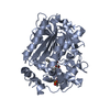
| ||||||||
| 2 | 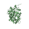
| ||||||||
| Unit cell |
|
-Components
| #1: Protein | Mass: 40513.633 Da / Num. of mol.: 2 Source method: isolated from a genetically manipulated source Source: (gene. exp.) Pseudonocardia thermophila (bacteria) / Gene: SAMN05443637_118146 / Production host: #2: Chemical |   Mass: 194.208 Da / Num. of mol.: 3 / Source method: obtained synthetically / Formula: C8H19O3P / Feature type: SUBJECT OF INVESTIGATION Mass: 194.208 Da / Num. of mol.: 3 / Source method: obtained synthetically / Formula: C8H19O3P / Feature type: SUBJECT OF INVESTIGATION#3: Chemical |   Mass: 176.169 Da / Num. of mol.: 2 / Source method: obtained synthetically / Formula: C10H8O3 / Feature type: SUBJECT OF INVESTIGATION Mass: 176.169 Da / Num. of mol.: 2 / Source method: obtained synthetically / Formula: C10H8O3 / Feature type: SUBJECT OF INVESTIGATION#4: Chemical | ChemComp-MG / |   Mass: 24.305 Da / Num. of mol.: 1 / Source method: obtained synthetically / Formula: Mg Mass: 24.305 Da / Num. of mol.: 1 / Source method: obtained synthetically / Formula: Mg#5: Water | ChemComp-HOH / |  Mass: 18.015 Da / Num. of mol.: 962 / Source method: isolated from a natural source / Formula: H2O Mass: 18.015 Da / Num. of mol.: 962 / Source method: isolated from a natural source / Formula: H2OHas ligand of interest | Y | Has protein modification | Y | |
|---|
-Experimental details
-Experiment
| Experiment | Method: X-RAY DIFFRACTION / Number of used crystals: 1 |
|---|
- Sample preparation
| Crystal | Density Matthews: 2.13 Å3/Da / Density % sol: 42.33 % |
|---|---|
| Crystal grow | Temperature: 285 K / Method: vapor diffusion, sitting drop Details: 0.2 M magnesium chloride, 0.1 M Tris pH 8.5, 30 % (w/v) PEG 4000, 2 mM methyl 4-methylumbelliferyl hexylphosphat |
-Data collection
| Diffraction | Mean temperature: 100 K / Serial crystal experiment: N |
|---|---|
| Diffraction source | Source: SYNCHROTRON / Site: ESRF  / Beamline: ID29 / Wavelength: 0.9762 Å / Beamline: ID29 / Wavelength: 0.9762 Å |
| Detector | Type: DECTRIS EIGER X 4M / Detector: PIXEL / Date: Sep 2, 2018 |
| Radiation | Protocol: SINGLE WAVELENGTH / Monochromatic (M) / Laue (L): M / Scattering type: x-ray |
| Radiation wavelength | Wavelength: 0.9762 Å / Relative weight: 1 |
| Reflection | Resolution: 1.81→43.39 Å / Num. obs: 63348 / % possible obs: 98.93 % / Redundancy: 4.9 % / CC1/2: 0.996 / Rmerge(I) obs: 0.117 / Net I/σ(I): 10.3 |
| Reflection shell | Resolution: 1.811→1.875 Å / Rmerge(I) obs: 0.555 / Num. unique obs: 6223 / CC1/2: 0.82 |
- Processing
| Software |
| ||||||||||||||||||||||||||||||||||||||||||||||||||||||||||||
|---|---|---|---|---|---|---|---|---|---|---|---|---|---|---|---|---|---|---|---|---|---|---|---|---|---|---|---|---|---|---|---|---|---|---|---|---|---|---|---|---|---|---|---|---|---|---|---|---|---|---|---|---|---|---|---|---|---|---|---|---|---|
| Refinement | Method to determine structure: MOLECULAR REPLACEMENT Starting model: 6Z68 Resolution: 1.81→43.39 Å / Cor.coef. Fo:Fc: 0.967 / Cor.coef. Fo:Fc free: 0.945 / SU B: 2.787 / SU ML: 0.083 / Cross valid method: THROUGHOUT / σ(F): 0 / ESU R: 0.122 / ESU R Free: 0.117 / Stereochemistry target values: MAXIMUM LIKELIHOOD Details: HYDROGENS HAVE BEEN ADDED IN THE RIDING POSITIONS U VALUES : REFINED INDIVIDUALLY
| ||||||||||||||||||||||||||||||||||||||||||||||||||||||||||||
| Solvent computation | Ion probe radii: 0.8 Å / Shrinkage radii: 0.8 Å / VDW probe radii: 1.2 Å / Solvent model: MASK | ||||||||||||||||||||||||||||||||||||||||||||||||||||||||||||
| Displacement parameters | Biso max: 79.78 Å2 / Biso mean: 15.82 Å2 / Biso min: 3.49 Å2
| ||||||||||||||||||||||||||||||||||||||||||||||||||||||||||||
| Refinement step | Cycle: final / Resolution: 1.81→43.39 Å
| ||||||||||||||||||||||||||||||||||||||||||||||||||||||||||||
| Refine LS restraints |
| ||||||||||||||||||||||||||||||||||||||||||||||||||||||||||||
| LS refinement shell | Resolution: 1.811→1.858 Å / Rfactor Rfree error: 0 / Total num. of bins used: 20
|
