 Movie
Movie Controller

[English] 日本語
 Yorodumi
- PDB-1s7h: Structural Genomics, 2.2A crystal structure of protein YKOF from ... -
+ Open data

- Basic information
| Entry | Database: PDB / ID: 1s7h | ||||||
|---|---|---|---|---|---|---|---|
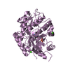
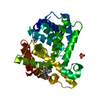
| Title | Structural Genomics, 2.2A crystal structure of protein YKOF from Bacillus subtilis | ||||||
 Components Components | ykoF | ||||||
 Keywords Keywords | STRUCTURAL GENOMICS / UNKNOWN FUNCTION / Protein YKOF / alpha-beta-beta-alpha sandwich / PSI / Protein Structure Initiative / Midwest Center for Structural Genomics / MCSG | ||||||
| Function / homology |  Function and homology information Function and homology information | ||||||
| Biological species |  | ||||||
| Method |  X-RAY DIFFRACTION / SYNCHROTRON / MAD / Resolution: 2.2 Å X-RAY DIFFRACTION / SYNCHROTRON / MAD / Resolution: 2.2 Å | ||||||
 Authors Authors | Zhang, R. / Lezondra, L. / Moy, S. / Dementieva, I. / Joachimiak, A. / Midwest Center for Structural Genomics (MCSG) | ||||||
 Citation Citation | Journal: To be Published Title: 2.2A crystal structure of protein YKOF from Bacillus subtilis Authors: Zhang, R. / Lezondra, L. / Moy, S. / Dementieva, I. / Joachimiak, A. | ||||||
| History |
|
- Structure visualization
| Structure viewer | Molecule: MolmilJmol/JSmol |
|---|
- Downloads & links
-Download
| PDBx/mmCIF format | 1s7h.cif.gz | 155.4 KB | Display | PDBx/mmCIF format |
|---|---|---|---|---|
| PDB format | pdb1s7h.ent.gz | 124 KB | Display | PDB format |
| PDBx/mmJSON format | 1s7h.json.gz | Tree view | PDBx/mmJSON format | |
| Others |  Other downloads Other downloads |
-Validation report
| Arichive directory | https://data.pdbj.org/pub/pdb/validation_reports/s7/1s7hftp://data.pdbj.org/pub/pdb/validation_reports/s7/1s7h | HTTPS FTP |
|---|
-Related structure data
| Similar structure data | |
|---|---|
| Other databases |


-Links
 PDBj
PDBj- Assembly
| Deposited unit | 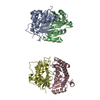
| ||||||||
|---|---|---|---|---|---|---|---|---|---|
| 1 | 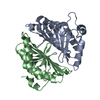
| ||||||||
| 2 | 
| ||||||||
| Unit cell |
| ||||||||
| Details | This protein exists as dimer. Molecules A and B, C and D represents two dimers in asymmeiric unit |
-Components
| #1: Protein | Mass: 22051.869 Da / Num. of mol.: 4 Source method: isolated from a genetically manipulated source Source: (gene. exp.) #2: Water | ChemComp-HOH / |  Mass: 18.015 Da / Num. of mol.: 372 / Source method: isolated from a natural source / Formula: H2O Mass: 18.015 Da / Num. of mol.: 372 / Source method: isolated from a natural source / Formula: H2O |
|---|
-Experimental details
-Experiment
| Experiment | Method: X-RAY DIFFRACTION / Number of used crystals: 1 |
|---|
- Sample preparation
| Crystal | Density Matthews: 2.53 Å3/Da / Density % sol: 50.99 % |
|---|---|
| Crystal grow | Temperature: 298 K / Method: vapor diffusion, hanging drop / pH: 8.5 Details: 0.2M MgCl2 Hexahydrate, 0.1M Tris , 25% PEG 3350, pH 8.5, VAPOR DIFFUSION, HANGING DROP, temperature 298K |
-Data collection
| Diffraction | Mean temperature: 100 K | |||||||||
|---|---|---|---|---|---|---|---|---|---|---|
| Diffraction source | Source: SYNCHROTRON / Site: APS  / Beamline: 19-ID / Wavelength: 0.9795, 0.9798 / Beamline: 19-ID / Wavelength: 0.9795, 0.9798 | |||||||||
| Detector | Type: SBC-2 / Detector: CCD / Date: Mar 10, 2003 / Details: mirrors | |||||||||
| Radiation | Monochromator: Si 111 channel / Protocol: MAD / Monochromatic (M) / Laue (L): M / Scattering type: x-ray | |||||||||
| Radiation wavelength |
| |||||||||
| Reflection | Resolution: 2.2→50 Å / Num. obs: 81572 / % possible obs: 99 % / Observed criterion σ(F): 4 / Observed criterion σ(I): 4 / Redundancy: 9.2 % / Biso Wilson estimate: 22.5 Å2 / Rmerge(I) obs: 0.089 / Net I/σ(I): 23.4 | |||||||||
| Reflection shell | Resolution: 2.2→2.25 Å / Redundancy: 7.8 % / Rmerge(I) obs: 0.4 / Mean I/σ(I) obs: 3.85 / Num. unique all: 2723 / % possible all: 95.2 |
- Processing
| Software |
| ||||||||||||||||||||
|---|---|---|---|---|---|---|---|---|---|---|---|---|---|---|---|---|---|---|---|---|---|
| Refinement | Method to determine structure: MAD / Resolution: 2.2→46.2 Å / Rfactor Rfree error: 0.005 / Data cutoff high absF: 320699.38 / Data cutoff low absF: 0 / Isotropic thermal model: RESTRAINED / Cross valid method: THROUGHOUT / σ(F): 0 / Stereochemistry target values: Engh & Huber / Details: Friedel's pairs were used in the refinement.
| ||||||||||||||||||||
| Solvent computation | Solvent model: FLAT MODEL / Bsol: 45.8256 Å2 / ksol: 0.321195 e/Å3 | ||||||||||||||||||||
| Displacement parameters | Biso mean: 35.3 Å2
| ||||||||||||||||||||
| Refine analyze |
| ||||||||||||||||||||
| Refinement step | Cycle: LAST / Resolution: 2.2→46.2 Å
| ||||||||||||||||||||
| Refine LS restraints |
| ||||||||||||||||||||
| LS refinement shell | Resolution: 2.2→2.34 Å / Rfactor Rfree error: 0.018 / Total num. of bins used: 6
| ||||||||||||||||||||
| Xplor file |
|
