 Movie
Movie Controller

[English] 日本語
 Yorodumi
- PDB-1qkr: Crystal structure of the vinculin tail and a pathway for activation -
+ Open data

- Basic information
| Entry | Database: PDB / ID: 1qkr | ||||||
|---|---|---|---|---|---|---|---|
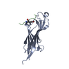
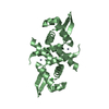
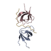
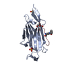
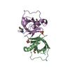
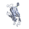
| Title | Crystal structure of the vinculin tail and a pathway for activation | ||||||
 Components Components | VINCULIN | ||||||
 Keywords Keywords | ACTIN CYTOSKELETON / CELL ADHESION / HELICAL BUNDLE / LIPID BINDING | ||||||
| Function / homology |  Function and homology information Function and homology informationmuscle tendon junction / Platelet degranulation / Smooth Muscle Contraction / regulation of protein localization to adherens junction / outer dense plaque of desmosome / inner dense plaque of desmosome / podosome ring / terminal web / epithelial cell-cell adhesion / zonula adherens ...muscle tendon junction / Platelet degranulation / Smooth Muscle Contraction / regulation of protein localization to adherens junction / outer dense plaque of desmosome / inner dense plaque of desmosome / podosome ring / terminal web / epithelial cell-cell adhesion / zonula adherens / Regulation of CDH1 Function / fascia adherens / MAP2K and MAPK activation / dystroglycan binding / alpha-catenin binding / muscle alpha-actinin binding / vinculin binding / cell-cell contact zone / apical junction assembly / costamere / regulation of establishment of endothelial barrier / adherens junction assembly / protein localization to cell surface / lamellipodium assembly / regulation of focal adhesion assembly / skeletal muscle myofibril / alpha-actinin binding / brush border / stress fiber / regulation of cell migration / Neutrophil degranulation / morphogenesis of an epithelium / adherens junction / neuromuscular junction / sarcolemma / beta-catenin binding / Z disc / cell-cell junction / actin filament binding / actin cytoskeleton / scaffold protein binding / cytoskeleton / cell adhesion / mitochondrial inner membrane / cadherin binding / focal adhesion / ubiquitin protein ligase binding / perinuclear region of cytoplasm / structural molecule activity / protein homodimerization activity / protein-containing complex / plasma membrane / cytoplasm Similarity search - Function | ||||||
| Biological species |  | ||||||
| Method |  X-RAY DIFFRACTION / SYNCHROTRON / SIRAS / Resolution: 1.8 Å X-RAY DIFFRACTION / SYNCHROTRON / SIRAS / Resolution: 1.8 Å | ||||||
 Authors Authors | Bakolitsa, C. / De Pereda, J.M. / Bagshaw, C.R. / Critchley, D.R. / Liddington, R.C. | ||||||
 Citation Citation | Journal: Cell(Cambridge,Mass.) / Year: 1999 Title: Crystal Structure of the Vinculin Tail and a Pathway for Activation Authors: Bakolitsa, C. / De Pereda, J.M. / Bagshaw, C.R. / Critchley, D.R. / Liddington, R.C. | ||||||
| History |
|
- Structure visualization
| Structure viewer | Molecule: MolmilJmol/JSmol |
|---|
- Downloads & links
-Download
| PDBx/mmCIF format | 1qkr.cif.gz | 89 KB | Display | PDBx/mmCIF format |
|---|---|---|---|---|
| PDB format | pdb1qkr.ent.gz | 68.8 KB | Display | PDB format |
| PDBx/mmJSON format | 1qkr.json.gz | Tree view | PDBx/mmJSON format | |
| Others |  Other downloads Other downloads |
-Validation report
| Arichive directory | https://data.pdbj.org/pub/pdb/validation_reports/qk/1qkrftp://data.pdbj.org/pub/pdb/validation_reports/qk/1qkr | HTTPS FTP |
|---|
-Related structure data
| Similar structure data |
|---|


-Links
 PDBj
PDBj
- Assembly
| Deposited unit | 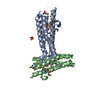
| ||||||||
|---|---|---|---|---|---|---|---|---|---|
| 1 | 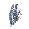
| ||||||||
| 2 | 
| ||||||||
| Unit cell |
|
-Components
| #1: Protein | Mass: 21629.486 Da / Num. of mol.: 2 / Fragment: C-TERMINAL DOMAIN Source method: isolated from a genetically manipulated source Source: (gene. exp.)  #2: Chemical |   Mass: 96.063 Da / Num. of mol.: 2 / Source method: obtained synthetically / Formula: SO4 Mass: 96.063 Da / Num. of mol.: 2 / Source method: obtained synthetically / Formula: SO4#3: Water | ChemComp-HOH / |  Mass: 18.015 Da / Num. of mol.: 316 / Source method: isolated from a natural source / Formula: H2O Mass: 18.015 Da / Num. of mol.: 316 / Source method: isolated from a natural source / Formula: H2OHas protein modification | Y | |
|---|
-Experimental details
-Experiment
| Experiment | Method: X-RAY DIFFRACTION |
|---|
- Sample preparation
| Crystal | Density Matthews: 2.03 Å3/Da / Density % sol: 44 % | ||||||||||||||||||||||||||||||||||||||||||||||||
|---|---|---|---|---|---|---|---|---|---|---|---|---|---|---|---|---|---|---|---|---|---|---|---|---|---|---|---|---|---|---|---|---|---|---|---|---|---|---|---|---|---|---|---|---|---|---|---|---|---|
| Crystal grow | pH: 5 Details: 25% (W/V) PEG 2000, 0.2 M (NH4)2SO4, 0.1 M CH3COO PH 5.0 | ||||||||||||||||||||||||||||||||||||||||||||||||
| Crystal | *PLUS Density % sol: 44 % | ||||||||||||||||||||||||||||||||||||||||||||||||
| Crystal grow | *PLUS Temperature: 22 ℃ / pH: 8 / Method: vapor diffusion, sitting drop | ||||||||||||||||||||||||||||||||||||||||||||||||
| Components of the solutions | *PLUS
|
-Data collection
| Diffraction | Mean temperature: 100 K |
|---|---|
| Diffraction source | Source: SYNCHROTRON / Site: SRS  / Beamline: PX9.6 / Wavelength: 1.488 / Beamline: PX9.6 / Wavelength: 1.488 |
| Detector | Type: MARRESEARCH / Detector: CCD |
| Radiation | Protocol: SINGLE WAVELENGTH / Monochromatic (M) / Laue (L): M / Scattering type: x-ray |
| Radiation wavelength | Wavelength: 1.488 Å / Relative weight: 1 |
| Reflection | Resolution: 1.8→10 Å / Num. obs: 30415 / % possible obs: 95.5 % / Redundancy: 2.6 % / Biso Wilson estimate: 18.4 Å2 / Rsym value: 0.064 / Net I/σ(I): 16.6 |
| Reflection shell | Resolution: 1.8→1.91 Å / Mean I/σ(I) obs: 3 / Rsym value: 0.27 / % possible all: 88.3 |
| Reflection | *PLUS Lowest resolution: 25 Å / % possible obs: 98.6 % / Rmerge(I) obs: 0.064 |
| Reflection shell | *PLUS Rmerge(I) obs: 0.27 / Mean I/σ(I) obs: 3 |
- Processing
| Software |
| ||||||||||||||||||||||||||||||||||||||||||||||||||||||||||||||||||||||||||||||||
|---|---|---|---|---|---|---|---|---|---|---|---|---|---|---|---|---|---|---|---|---|---|---|---|---|---|---|---|---|---|---|---|---|---|---|---|---|---|---|---|---|---|---|---|---|---|---|---|---|---|---|---|---|---|---|---|---|---|---|---|---|---|---|---|---|---|---|---|---|---|---|---|---|---|---|---|---|---|---|---|---|---|
| Refinement | Method to determine structure: SIRAS / Resolution: 1.8→10 Å / Rfactor Rfree error: 0.006 / Isotropic thermal model: RESTRAINED / Cross valid method: THROUGHOUT / σ(F): 0
| ||||||||||||||||||||||||||||||||||||||||||||||||||||||||||||||||||||||||||||||||
| Solvent computation | Solvent model: FLAT MODEL / Bsol: 70.2752 Å2 / ksol: 0.426906 e/Å3 | ||||||||||||||||||||||||||||||||||||||||||||||||||||||||||||||||||||||||||||||||
| Displacement parameters | Biso mean: 25.9 Å2
| ||||||||||||||||||||||||||||||||||||||||||||||||||||||||||||||||||||||||||||||||
| Refine analyze |
| ||||||||||||||||||||||||||||||||||||||||||||||||||||||||||||||||||||||||||||||||
| Refinement step | Cycle: LAST / Resolution: 1.8→10 Å
| ||||||||||||||||||||||||||||||||||||||||||||||||||||||||||||||||||||||||||||||||
| Refine LS restraints |
| ||||||||||||||||||||||||||||||||||||||||||||||||||||||||||||||||||||||||||||||||
| LS refinement shell | Resolution: 1.8→1.91 Å / Rfactor Rfree error: 0.019 / Total num. of bins used: 6
| ||||||||||||||||||||||||||||||||||||||||||||||||||||||||||||||||||||||||||||||||
| Xplor file |
| ||||||||||||||||||||||||||||||||||||||||||||||||||||||||||||||||||||||||||||||||
| Software | *PLUS Name: CNS / Version: 0.5 / Classification: refinement | ||||||||||||||||||||||||||||||||||||||||||||||||||||||||||||||||||||||||||||||||
| Refinement | *PLUS Rfactor obs: 0.2 / Rfactor Rwork: 0.2 | ||||||||||||||||||||||||||||||||||||||||||||||||||||||||||||||||||||||||||||||||
| Solvent computation | *PLUS | ||||||||||||||||||||||||||||||||||||||||||||||||||||||||||||||||||||||||||||||||
| Displacement parameters | *PLUS | ||||||||||||||||||||||||||||||||||||||||||||||||||||||||||||||||||||||||||||||||
| Refine LS restraints | *PLUS
| ||||||||||||||||||||||||||||||||||||||||||||||||||||||||||||||||||||||||||||||||
| LS refinement shell | *PLUS Rfactor Rfree: 0.28 |
