 Movie
Movie Controller

[English] 日本語
 Yorodumi
- PDB-5sxp: STRUCTURAL BASIS FOR THE INTERACTION BETWEEN ITCH PRR AND BETA-PIX -
+ Open data

- Basic information
| Entry | Database: PDB / ID: 5sxp | ||||||
|---|---|---|---|---|---|---|---|
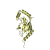
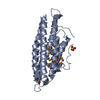
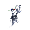
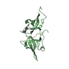
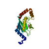
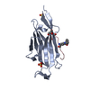
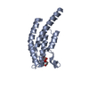
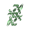
| Title | STRUCTURAL BASIS FOR THE INTERACTION BETWEEN ITCH PRR AND BETA-PIX | ||||||
 Components Components |
| ||||||
 Keywords Keywords | SIGNALING PROTEIN/LIGASE / SH3 DOMAIN PEPTIDE LIGAND COMPLEX / LIGASE / SIGNALING PROTEIN-LIGASE complex | ||||||
| Function / homology |  Function and homology information Function and homology informationnegative regulation of microtubule nucleation / regulation of protein deubiquitination / negative regulation of cytoplasmic pattern recognition receptor signaling pathway / ubiquitin-like protein transferase activity / nucleotide-binding domain, leucine rich repeat containing receptor signaling pathway / negative regulation of defense response to virus / protein K29-linked ubiquitination / T cell anergy / CXCR chemokine receptor binding / regulation of necroptotic process ...negative regulation of microtubule nucleation / regulation of protein deubiquitination / negative regulation of cytoplasmic pattern recognition receptor signaling pathway / ubiquitin-like protein transferase activity / nucleotide-binding domain, leucine rich repeat containing receptor signaling pathway / negative regulation of defense response to virus / protein K29-linked ubiquitination / T cell anergy / CXCR chemokine receptor binding / regulation of necroptotic process / protein branched polyubiquitination / positive regulation of T cell anergy / CD4-positive, alpha-beta T cell proliferation / negative regulation of CD4-positive, alpha-beta T cell proliferation / focal adhesion assembly / HECT-type E3 ubiquitin transferase / gamma-tubulin binding / arrestin family protein binding / positive regulation of lamellipodium morphogenesis / Ephrin signaling / regulation of hematopoietic stem cell differentiation / positive regulation of fibroblast migration / negative regulation of JNK cascade / lamellipodium assembly / positive regulation of receptor catabolic process / RHOV GTPase cycle / NRAGE signals death through JNK / ubiquitin-ubiquitin ligase activity / mitotic spindle pole / negative regulation of type I interferon production / Activation of RAC1 downstream of NMDARs / RHOJ GTPase cycle / ligase activity / RHOQ GTPase cycle / Golgi organization / RHOU GTPase cycle / CDC42 GTPase cycle / ubiquitin-like protein ligase binding / RHOA GTPase cycle / ephrin receptor signaling pathway / ribonucleoprotein complex binding / protein K63-linked ubiquitination / protein monoubiquitination / protein autoubiquitination / Rho protein signal transduction / ruffle / RAC1 GTPase cycle / protein K48-linked ubiquitination / positive regulation of substrate adhesion-dependent cell spreading / guanyl-nucleotide exchange factor activity / Downregulation of ERBB4 signaling / Activated NOTCH1 Transmits Signal to the Nucleus / negative regulation of canonical NF-kappaB signal transduction / Negative regulators of DDX58/IFIH1 signaling / regulation of cell growth / EGFR downregulation / NOD1/2 Signaling Pathway / Degradation of GLI1 by the proteasome / receptor internalization / Hedgehog 'on' state / Regulation of necroptotic cell death / ubiquitin-protein transferase activity / SARS-CoV-1 activates/modulates innate immune responses / positive regulation of protein catabolic process / ubiquitin protein ligase activity / nervous system development / lamellipodium / Antigen processing: Ubiquitination & Proteasome degradation / G alpha (12/13) signalling events / RUNX1 regulates transcription of genes involved in differentiation of HSCs / cytoplasmic vesicle / early endosome membrane / cell cortex / defense response to virus / ubiquitin-dependent protein catabolic process / proteasome-mediated ubiquitin-dependent protein catabolic process / neuron projection / postsynapse / protein ubiquitination / positive regulation of apoptotic process / inflammatory response / innate immune response / focal adhesion / neuronal cell body / apoptotic process / symbiont entry into host cell / centrosome / protein kinase binding / negative regulation of apoptotic process / signal transduction / protein-containing complex / mitochondrion / extracellular exosome / nucleoplasm / membrane / plasma membrane / cytoplasm / cytosol Similarity search - Function | ||||||
| Biological species |  Homo sapiens (human) Homo sapiens (human) | ||||||
| Method |  X-RAY DIFFRACTION / SYNCHROTRON / MOLECULAR REPLACEMENT / Resolution: 1.65 Å X-RAY DIFFRACTION / SYNCHROTRON / MOLECULAR REPLACEMENT / Resolution: 1.65 Å | ||||||
 Authors Authors | Cappadocia, L. / Desrochers, G. / Lussier-Price, M. / Angers, A. / Omichinski, J.G. | ||||||
| Funding support |  Canada, 1items Canada, 1items
| ||||||
 Citation Citation | Journal: J. Biol. Chem. / Year: 2017 Title: Molecular basis of interactions between SH3 domain-containing proteins and the proline-rich region of the ubiquitin ligase Itch. Authors: Desrochers, G. / Cappadocia, L. / Lussier-Price, M. / Ton, A.T. / Ayoubi, R. / Serohijos, A. / Omichinski, J.G. / Angers, A. | ||||||
| History |
|
- Structure visualization
| Structure viewer | Molecule: MolmilJmol/JSmol |
|---|
- Downloads & links
-Download
| PDBx/mmCIF format | 5sxp.cif.gz | 181 KB | Display | PDBx/mmCIF format |
|---|---|---|---|---|
| PDB format | pdb5sxp.ent.gz | 147 KB | Display | PDB format |
| PDBx/mmJSON format | 5sxp.json.gz | Tree view | PDBx/mmJSON format | |
| Others |  Other downloads Other downloads |
-Validation report
| Arichive directory | https://data.pdbj.org/pub/pdb/validation_reports/sx/5sxpftp://data.pdbj.org/pub/pdb/validation_reports/sx/5sxp | HTTPS FTP |
|---|
-Related structure data
| Related structure data | 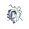 2p4rS S: Starting model for refinement |
|---|---|
| Similar structure data |


-Links
 PDBj
PDBj


- Assembly
| Deposited unit | 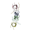
| ||||||||
|---|---|---|---|---|---|---|---|---|---|
| 1 | 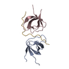
| ||||||||
| 2 | 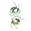
| ||||||||
| Unit cell |
|
-Components
| #1: Protein | Mass: 7094.742 Da / Num. of mol.: 4 / Fragment: UNP residues 183-243 Source method: isolated from a genetically manipulated source Source: (gene. exp.) Homo sapiens (human)Gene: ARHGEF7, COOL1, KIAA0142, P85SPR, PAK3BP, PIXB, Nbla10314 Plasmid: pGEX-4T1 / Production host:  #2: Protein/peptide | Mass: 2816.185 Da / Num. of mol.: 2 / Fragment: UNP residues 249-269 Source method: isolated from a genetically manipulated source Source: (gene. exp.) Homo sapiens (human) / Gene: ITCH / Plasmid: pGEX-4T1 / Production host: References: UniProt: Q96J02, Ligases; Forming carbon-nitrogen bonds; Acid-amino-acid ligases (peptide synthases) #3: Water | ChemComp-HOH / |  Mass: 18.015 Da / Num. of mol.: 383 / Source method: isolated from a natural source / Formula: H2O Mass: 18.015 Da / Num. of mol.: 383 / Source method: isolated from a natural source / Formula: H2O |
|---|
-Experimental details
-Experiment
| Experiment | Method: X-RAY DIFFRACTION / Number of used crystals: 1 |
|---|
- Sample preparation
| Crystal | Density Matthews: 2.14 Å3/Da / Density % sol: 42.58 % |
|---|---|
| Crystal grow | Temperature: 293 K / Method: vapor diffusion, hanging drop / Details: 100 mM MIB buffer pH 5.0 and 25% PEG1500 |
-Data collection
| Diffraction | Mean temperature: 100 K |
|---|---|
| Diffraction source | Source: SYNCHROTRON / Site: NSLS  / Beamline: X25 / Wavelength: 1.1 Å / Beamline: X25 / Wavelength: 1.1 Å |
| Detector | Type: DECTRIS PILATUS 6M / Detector: PIXEL / Date: Feb 18, 2013 |
| Radiation | Monochromator: Si(111) / Protocol: SINGLE WAVELENGTH / Monochromatic (M) / Laue (L): M / Scattering type: x-ray |
| Radiation wavelength | Wavelength: 1.1 Å / Relative weight: 1 |
| Reflection | Resolution: 1.65→50 Å / Num. obs: 32525 / % possible obs: 95.7 % / Observed criterion σ(F): -3 / Redundancy: 1.8 % / Rmerge(I) obs: 0.021 / Net I/σ(I): 18.2 |
| Reflection shell | Resolution: 1.65→1.74 Å / Redundancy: 1.8 % / Rmerge(I) obs: 0.069 / Mean I/σ(I) obs: 6.9 / % possible all: 93.2 |
- Processing
| Software |
| |||||||||||||||||||||||||||||||||||||||||||||||||||||||||||||||||||||||||||||
|---|---|---|---|---|---|---|---|---|---|---|---|---|---|---|---|---|---|---|---|---|---|---|---|---|---|---|---|---|---|---|---|---|---|---|---|---|---|---|---|---|---|---|---|---|---|---|---|---|---|---|---|---|---|---|---|---|---|---|---|---|---|---|---|---|---|---|---|---|---|---|---|---|---|---|---|---|---|---|
| Refinement | Method to determine structure: MOLECULAR REPLACEMENT Starting model: 2P4R Resolution: 1.65→41.847 Å / SU ML: 0.12 / Cross valid method: FREE R-VALUE / σ(F): 1.98 / Phase error: 16.37 / Stereochemistry target values: ML
| |||||||||||||||||||||||||||||||||||||||||||||||||||||||||||||||||||||||||||||
| Solvent computation | Shrinkage radii: 0.9 Å / VDW probe radii: 1.11 Å / Solvent model: FLAT BULK SOLVENT MODEL | |||||||||||||||||||||||||||||||||||||||||||||||||||||||||||||||||||||||||||||
| Refinement step | Cycle: LAST / Resolution: 1.65→41.847 Å
| |||||||||||||||||||||||||||||||||||||||||||||||||||||||||||||||||||||||||||||
| Refine LS restraints |
| |||||||||||||||||||||||||||||||||||||||||||||||||||||||||||||||||||||||||||||
| LS refinement shell |
|
