 Movie
Movie Controller

[English] 日本語
 Yorodumi
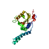
- PDB-5yy9: Crystal structure of Tandem Tudor Domain of human UHRF1 in comple... -
+ Open data

- Basic information
| Entry | Database: PDB / ID: 5yy9 | ||||||
|---|---|---|---|---|---|---|---|
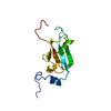
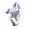
| Title | Crystal structure of Tandem Tudor Domain of human UHRF1 in complex with LIG1-K126me3 | ||||||
 Components Components |
| ||||||
 Keywords Keywords | TRANSFERASE / Maintenance of DNA methylation | ||||||
| Function / homology |  Function and homology information Function and homology informationOkazaki fragment processing involved in mitotic DNA replication / histone H3K18 ubiquitin ligase activity / histone H3K14 ubiquitin ligase activity / histone H3 ubiquitin ligase activity / histone H3K23 ubiquitin ligase activity / DNA ligase activity / chromosomal DNA methylation maintenance following DNA replication / DNA damage sensor activity / DNA ligase (ATP) / DNA ligase (ATP) activity ...Okazaki fragment processing involved in mitotic DNA replication / histone H3K18 ubiquitin ligase activity / histone H3K14 ubiquitin ligase activity / histone H3 ubiquitin ligase activity / histone H3K23 ubiquitin ligase activity / DNA ligase activity / chromosomal DNA methylation maintenance following DNA replication / DNA damage sensor activity / DNA ligase (ATP) / DNA ligase (ATP) activity / telomere maintenance via semi-conservative replication / hemi-methylated DNA-binding / homologous recombination / Regulation of MITF-M-dependent genes involved in DNA replication, damage repair and senescence / regulation of epithelial cell proliferation / Processive synthesis on the lagging strand / lagging strand elongation / Mismatch repair (MMR) directed by MSH2:MSH3 (MutSbeta) / Mismatch repair (MMR) directed by MSH2:MSH6 (MutSalpha) / methyl-CpG binding / Processive synthesis on the C-strand of the telomere / histone H3K9me2/3 reader activity / negative regulation of gene expression via chromosomal CpG island methylation / DNA biosynthetic process / Early Phase of HIV Life Cycle / positive regulation of protein metabolic process / POLB-Dependent Long Patch Base Excision Repair / PCNA-Dependent Long Patch Base Excision Repair / mitotic spindle assembly / anatomical structure morphogenesis / mismatch repair / base-excision repair, gap-filling / protein autoubiquitination / cis-regulatory region sequence-specific DNA binding / protein localization to chromatin / heterochromatin / epigenetic regulation of gene expression / DNA methylation / Chromatin modifications during the maternal to zygotic transition (MZT) / replication fork / Gap-filling DNA repair synthesis and ligation in GG-NER / euchromatin / base-excision repair / RING-type E3 ubiquitin transferase / double-strand break repair via homologous recombination / spindle / nuclear matrix / ubiquitin-protein transferase activity / Gap-filling DNA repair synthesis and ligation in TC-NER / ubiquitin protein ligase activity / heterochromatin formation / DNA recombination / histone binding / nucleic acid binding / ubiquitin-dependent protein catabolic process / protein ubiquitination / cell division / DNA repair / DNA damage response / chromatin / negative regulation of transcription by RNA polymerase II / positive regulation of transcription by RNA polymerase II / DNA binding / zinc ion binding / nucleoplasm / ATP binding / metal ion binding / identical protein binding / nucleus Similarity search - Function | ||||||
| Biological species |  Homo sapiens (human) Homo sapiens (human)synthetic construct (others) | ||||||
| Method |  X-RAY DIFFRACTION / SYNCHROTRON / MOLECULAR REPLACEMENT / Resolution: 2.653 Å X-RAY DIFFRACTION / SYNCHROTRON / MOLECULAR REPLACEMENT / Resolution: 2.653 Å | ||||||
 Authors Authors | Kori, S. / Defossez, P.A. / Arita, K. | ||||||
| Funding support |  Japan, 1items Japan, 1items
| ||||||
 Citation Citation | Journal: Structure / Year: 2019 Title: Structure of the UHRF1 Tandem Tudor Domain Bound to a Methylated Non-histone Protein, LIG1, Reveals Rules for Binding and Regulation. Authors: Kori, S. / Ferry, L. / Matano, S. / Jimenji, T. / Kodera, N. / Tsusaka, T. / Matsumura, R. / Oda, T. / Sato, M. / Dohmae, N. / Ando, T. / Shinkai, Y. / Defossez, P.A. / Arita, K. | ||||||
| History |
|
- Structure visualization
| Structure viewer | Molecule: MolmilJmol/JSmol |
|---|
- Downloads & links
-Download
| PDBx/mmCIF format | 5yy9.cif.gz | 75.4 KB | Display | PDBx/mmCIF format |
|---|---|---|---|---|
| PDB format | pdb5yy9.ent.gz | 53.8 KB | Display | PDB format |
| PDBx/mmJSON format | 5yy9.json.gz | Tree view | PDBx/mmJSON format | |
| Others |  Other downloads Other downloads |
-Validation report
| Arichive directory | https://data.pdbj.org/pub/pdb/validation_reports/yy/5yy9ftp://data.pdbj.org/pub/pdb/validation_reports/yy/5yy9 | HTTPS FTP |
|---|
-Related structure data
| Related structure data | 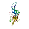 5yyaC 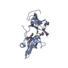 3db3S S: Starting model for refinement C: citing same article ( |
|---|---|
| Similar structure data |


-Links
 PDBj
PDBj


- Assembly
| Deposited unit | 
| ||||||||
|---|---|---|---|---|---|---|---|---|---|
| 1 | 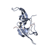
| ||||||||
| 2 | 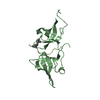
| ||||||||
| Unit cell |
|
-Components
| #1: Protein | Mass: 18162.207 Da / Num. of mol.: 2 / Fragment: Tandem Tudor Domain Source method: isolated from a genetically manipulated source Source: (gene. exp.) Homo sapiens (human) / Gene: UHRF1, ICBP90, NP95, RNF106 / Production host:  References: UniProt: Q96T88, RING-type E3 ubiquitin transferase #2: Protein/peptide | Mass: 1640.072 Da / Num. of mol.: 2 / Source method: obtained synthetically / Source: (synth.) synthetic construct (others) / References: UniProt: P18858*PLUS #3: Water | ChemComp-HOH / |  Mass: 18.015 Da / Num. of mol.: 24 / Source method: isolated from a natural source / Formula: H2O Mass: 18.015 Da / Num. of mol.: 24 / Source method: isolated from a natural source / Formula: H2O |
|---|
-Experimental details
-Experiment
| Experiment | Method: X-RAY DIFFRACTION / Number of used crystals: 1 |
|---|
- Sample preparation
| Crystal | Density Matthews: 2.1 Å3/Da / Density % sol: 41.32 % |
|---|---|
| Crystal grow | Temperature: 293 K / Method: vapor diffusion, hanging drop / pH: 7 Details: 00 mM Tris-HCl (pH 7.0), 200 mM tri-potassium phosphate and 20% (w/v) PEG 3350 |
-Data collection
| Diffraction | Mean temperature: 100 K |
|---|---|
| Diffraction source | Source: SYNCHROTRON / Site: Photon Factory / Beamline: BL-17A / Wavelength: 0.98 Å |
| Detector | Type: DECTRIS PILATUS3 6M / Detector: PIXEL / Date: May 1, 2017 |
| Radiation | Protocol: SINGLE WAVELENGTH / Monochromatic (M) / Laue (L): M / Scattering type: x-ray |
| Radiation wavelength | Wavelength: 0.98 Å / Relative weight: 1 |
| Reflection | Resolution: 2.65→48.67 Å / Num. obs: 40019 / % possible obs: 97 % / Redundancy: 3.8 % / CC1/2: 0.99 / Rmerge(I) obs: 0.12 / Net I/σ(I): 8.5 |
| Reflection shell | Resolution: 2.65→2.78 Å / Redundancy: 3.8 % / Rmerge(I) obs: 0.7 / Mean I/σ(I) obs: 2.4 / CC1/2: 0.95 / % possible all: 95.4 |
- Processing
| Software |
| ||||||||||||||||||||||||||||||||||||||||||||||||||||||||
|---|---|---|---|---|---|---|---|---|---|---|---|---|---|---|---|---|---|---|---|---|---|---|---|---|---|---|---|---|---|---|---|---|---|---|---|---|---|---|---|---|---|---|---|---|---|---|---|---|---|---|---|---|---|---|---|---|---|
| Refinement | Method to determine structure: MOLECULAR REPLACEMENT Starting model: 3DB3 Resolution: 2.653→40.228 Å / SU ML: 0.35 / Cross valid method: FREE R-VALUE / σ(F): 1.36 / Phase error: 30.67
| ||||||||||||||||||||||||||||||||||||||||||||||||||||||||
| Solvent computation | Shrinkage radii: 0.9 Å / VDW probe radii: 1.11 Å | ||||||||||||||||||||||||||||||||||||||||||||||||||||||||
| Refinement step | Cycle: LAST / Resolution: 2.653→40.228 Å
| ||||||||||||||||||||||||||||||||||||||||||||||||||||||||
| Refine LS restraints |
| ||||||||||||||||||||||||||||||||||||||||||||||||||||||||
| LS refinement shell |
|
