 Movie
Movie Controller


+ Open data

- Basic information
| Entry | Database: PDB / ID: 5hb5 | ||||||||||||||||||||||||||||||
|---|---|---|---|---|---|---|---|---|---|---|---|---|---|---|---|---|---|---|---|---|---|---|---|---|---|---|---|---|---|---|---|
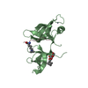
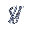
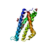
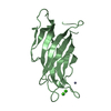
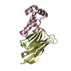
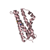
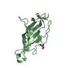
| Title | Crystal structure of Chaetomium thermophilum Nup145N APD | ||||||||||||||||||||||||||||||
 Components Components | Nucleoporin NUP145 | ||||||||||||||||||||||||||||||
 Keywords Keywords | TRANSPORT PROTEIN / Nucleocytoplasmic transport / Protein transport | ||||||||||||||||||||||||||||||
| Function / homology |  Function and homology information Function and homology informationtelomere tethering at nuclear periphery / nuclear pore cytoplasmic filaments / post-transcriptional tethering of RNA polymerase II gene DNA at nuclear periphery / structural constituent of nuclear pore / RNA export from nucleus / nuclear localization sequence binding / Hydrolases; Acting on peptide bonds (peptidases); Serine endopeptidases / mRNA transport / protein import into nucleus / nuclear membrane ...telomere tethering at nuclear periphery / nuclear pore cytoplasmic filaments / post-transcriptional tethering of RNA polymerase II gene DNA at nuclear periphery / structural constituent of nuclear pore / RNA export from nucleus / nuclear localization sequence binding / Hydrolases; Acting on peptide bonds (peptidases); Serine endopeptidases / mRNA transport / protein import into nucleus / nuclear membrane / hydrolase activity / RNA binding Similarity search - Function | ||||||||||||||||||||||||||||||
| Biological species |  Chaetomium thermophilum (fungus) Chaetomium thermophilum (fungus) | ||||||||||||||||||||||||||||||
| Method |  X-RAY DIFFRACTION / SYNCHROTRON / Resolution: 1.5 Å X-RAY DIFFRACTION / SYNCHROTRON / Resolution: 1.5 Å | ||||||||||||||||||||||||||||||
 Authors Authors | Lin, D.H. / Stuwe, T. / Hoelz, A. | ||||||||||||||||||||||||||||||
| Funding support |  United States, United States,  Germany, Germany,  China, 9items China, 9items
| ||||||||||||||||||||||||||||||
 Citation Citation | Journal: Science / Year: 2016 Title: Architecture of the symmetric core of the nuclear pore. Authors: Lin, D.H. / Stuwe, T. / Schilbach, S. / Rundlet, E.J. / Perriches, T. / Mobbs, G. / Fan, Y. / Thierbach, K. / Huber, F.M. / Collins, L.N. / Davenport, A.M. / Jeon, Y.E. / Hoelz, A. | ||||||||||||||||||||||||||||||
| History |
|
- Structure visualization
| Structure viewer | Molecule: MolmilJmol/JSmol |
|---|
- Downloads & links
-Download
| PDBx/mmCIF format | 5hb5.cif.gz | 185.5 KB | Display | PDBx/mmCIF format |
|---|---|---|---|---|
| PDB format | pdb5hb5.ent.gz | 153.1 KB | Display | PDB format |
| PDBx/mmJSON format | 5hb5.json.gz | Tree view | PDBx/mmJSON format | |
| Others |  Other downloads Other downloads |
-Validation report
| Arichive directory | https://data.pdbj.org/pub/pdb/validation_reports/hb/5hb5ftp://data.pdbj.org/pub/pdb/validation_reports/hb/5hb5 | HTTPS FTP |
|---|
-Related structure data
| Related structure data | 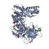 5haxC  5hayC  5hazC 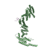 5hb0C  5hb1C  5hb2C  5hb3C  5hb4C  5hb6C  5hb7C  5hb8C C: citing same article ( |
|---|---|
| Similar structure data |


-Links
 PDBj
PDBj


- Assembly
| Deposited unit | 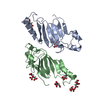
| ||||||||
|---|---|---|---|---|---|---|---|---|---|
| 1 | 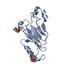
| ||||||||
| 2 | 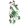
| ||||||||
| Unit cell |
|
-Components
| #1: Protein | Mass: 15693.921 Da / Num. of mol.: 2 Source method: isolated from a genetically manipulated source Source: (gene. exp.) Chaetomium thermophilum (strain DSM 1495 / CBS 144.50 / IMI 039719) (fungus)Strain: DSM 1495 / CBS 144.50 / IMI 039719 / Gene: NUP145, CTHT_0042590 / Production host:  References: UniProt: G0SAK3, Hydrolases; Acting on peptide bonds (peptidases); Serine endopeptidases #2: Chemical | ChemComp-FLC /   Mass: 189.100 Da / Num. of mol.: 7 / Source method: obtained synthetically / Formula: C6H5O7 Mass: 189.100 Da / Num. of mol.: 7 / Source method: obtained synthetically / Formula: C6H5O7#3: Water | ChemComp-HOH / |  Mass: 18.015 Da / Num. of mol.: 323 / Source method: isolated from a natural source / Formula: H2O Mass: 18.015 Da / Num. of mol.: 323 / Source method: isolated from a natural source / Formula: H2O |
|---|
-Experimental details
-Experiment
| Experiment | Method: X-RAY DIFFRACTION |
|---|
- Sample preparation
| Crystal | Density Matthews: 1.97 Å3/Da / Density % sol: 37.68 % |
|---|---|
| Crystal grow | Temperature: 294 K / Method: vapor diffusion, hanging drop / Details: 0.1 M citric acid (pH 3.0), 25 % (w/v) PEG 3350 |
-Data collection
| Diffraction | Mean temperature: 100 K |
|---|---|
| Diffraction source | Source: SYNCHROTRON / Site: SSRL / Beamline: BL12-2 / Wavelength: 0.9795 Å |
| Detector | Type: DECTRIS PILATUS 6M / Detector: PIXEL / Date: Apr 20, 2012 |
| Radiation | Protocol: SINGLE WAVELENGTH / Monochromatic (M) / Laue (L): M / Scattering type: x-ray |
| Radiation wavelength | Wavelength: 0.9795 Å / Relative weight: 1 |
| Reflection | Resolution: 1.5→30 Å / Num. obs: 38743 / % possible obs: 97.2 % / Redundancy: 6.6 % / Net I/σ(I): 12.4 |
- Processing
| Software |
| |||||||||||||||||||||||||||||||||||||||||||||||||||||||||||||||||||||||||||||||||||||||||||||||||||||||||
|---|---|---|---|---|---|---|---|---|---|---|---|---|---|---|---|---|---|---|---|---|---|---|---|---|---|---|---|---|---|---|---|---|---|---|---|---|---|---|---|---|---|---|---|---|---|---|---|---|---|---|---|---|---|---|---|---|---|---|---|---|---|---|---|---|---|---|---|---|---|---|---|---|---|---|---|---|---|---|---|---|---|---|---|---|---|---|---|---|---|---|---|---|---|---|---|---|---|---|---|---|---|---|---|---|---|---|
| Refinement | Resolution: 1.5→27.663 Å / SU ML: 0.15 / Cross valid method: THROUGHOUT / σ(F): 1.36 / Phase error: 24.15 / Stereochemistry target values: ML
| |||||||||||||||||||||||||||||||||||||||||||||||||||||||||||||||||||||||||||||||||||||||||||||||||||||||||
| Solvent computation | Shrinkage radii: 0.9 Å / VDW probe radii: 1.11 Å / Solvent model: FLAT BULK SOLVENT MODEL | |||||||||||||||||||||||||||||||||||||||||||||||||||||||||||||||||||||||||||||||||||||||||||||||||||||||||
| Refinement step | Cycle: LAST / Resolution: 1.5→27.663 Å
| |||||||||||||||||||||||||||||||||||||||||||||||||||||||||||||||||||||||||||||||||||||||||||||||||||||||||
| Refine LS restraints |
| |||||||||||||||||||||||||||||||||||||||||||||||||||||||||||||||||||||||||||||||||||||||||||||||||||||||||
| LS refinement shell |
|
