 Movie
Movie Controller


+ Open data

- Basic information
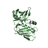
| Entry | Database: PDB / ID: 3a5p | ||||||
|---|---|---|---|---|---|---|---|
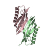
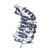
| Title | Crystal structure of hemagglutinin | ||||||
 Components Components | Haemagglutinin I | ||||||
 Keywords Keywords | SUGAR BINDING PROTEIN / lectin | ||||||
| Function / homology | Tumour Suppressor Smad4 - #70 / Hemagglutinin I / Hemagglutinin I / Tumour Suppressor Smad4 / Sandwich / Mainly Beta / Haemagglutinin I Function and homology information Function and homology information | ||||||
| Biological species |  Physarum polycephalum (eukaryote) Physarum polycephalum (eukaryote) | ||||||
| Method |  X-RAY DIFFRACTION / MOLECULAR REPLACEMENT / Resolution: 1.82 Å X-RAY DIFFRACTION / MOLECULAR REPLACEMENT / Resolution: 1.82 Å | ||||||
 Authors Authors | Watanabe, N. / Sakai, N. / Nakamura, T. / Nabeshima, Y. / Kouno, T. / Mizuguchi, M. / Kawano, K. | ||||||
 Citation Citation | Journal: J.Mol.Biol. / Year: 2011 Title: The Structure of Physarum polycephalum hemagglutinin I suggests a minimal carbohydrate recognition domain of legume lectin fold Authors: Kouno, T. / Watanabe, N. / Sakai, N. / Nakamura, T. / Nabeshima, Y. / Morita, M. / Mizuguchi, M. / Aizawa, T. / Demura, M. / Imanaka, T. / Tanaka, I. / Kawano, K. | ||||||
| History |
|
- Structure visualization
| Structure viewer | Molecule: MolmilJmol/JSmol |
|---|
- Downloads & links
-Download
| PDBx/mmCIF format | 3a5p.cif.gz | 107.5 KB | Display | PDBx/mmCIF format |
|---|---|---|---|---|
| PDB format | pdb3a5p.ent.gz | 83.1 KB | Display | PDB format |
| PDBx/mmJSON format | 3a5p.json.gz | Tree view | PDBx/mmJSON format | |
| Others |  Other downloads Other downloads |
-Validation report
| Arichive directory | https://data.pdbj.org/pub/pdb/validation_reports/a5/3a5pftp://data.pdbj.org/pub/pdb/validation_reports/a5/3a5p | HTTPS FTP |
|---|
-Related structure data
| Similar structure data |
|---|


-Links
 PDBj
PDBj- Assembly
| Deposited unit | 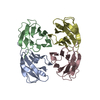
| ||||||||
|---|---|---|---|---|---|---|---|---|---|
| 1 | 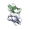
| ||||||||
| 2 | 
| ||||||||
| Unit cell |
|
-Components
| #1: Protein | Mass: 10900.101 Da / Num. of mol.: 4 Source method: isolated from a genetically manipulated source Source: (gene. exp.) Physarum polycephalum (eukaryote) / Plasmid: pET3a / Production host:  #2: Water | ChemComp-HOH / |  Mass: 18.015 Da / Num. of mol.: 858 / Source method: isolated from a natural source / Formula: H2O Mass: 18.015 Da / Num. of mol.: 858 / Source method: isolated from a natural source / Formula: H2O |
|---|
-Experimental details
-Experiment
| Experiment | Method: X-RAY DIFFRACTION / Number of used crystals: 1 |
|---|
- Sample preparation
| Crystal | Density Matthews: 2.5 Å3/Da / Density % sol: 50.8 % |
|---|---|
| Crystal grow | Temperature: 293 K / Method: vapor diffusion, hanging drop / pH: 6.5 Details: 15% PEG3350, 150mM NaI, pH 6.5, vapour diffusion, hanging drop, temperature 293K |
-Data collection
| Diffraction | Mean temperature: 100 K |
|---|---|
| Diffraction source | Source: ROTATING ANODE / Type: RIGAKU FR-E SUPERBRIGHT / Wavelength: 1.502 Å |
| Detector | Type: RIGAKU RAXIS VII / Detector: IMAGE PLATE / Date: Sep 8, 2006 / Details: mirror |
| Radiation | Protocol: SINGLE WAVELENGTH / Scattering type: x-ray |
| Radiation wavelength | Wavelength: 1.502 Å / Relative weight: 1 |
| Reflection | Resolution: 1.82→50 Å / Num. obs: 40006 / % possible obs: 99.8 % / Redundancy: 2.5 % / Biso Wilson estimate: 21.97 Å2 / Rsym value: 0.03 / Net I/σ(I): 49.4 |
| Reflection shell | Resolution: 1.82→1.89 Å / Redundancy: 2.3 % / Mean I/σ(I) obs: 11 / Rsym value: 0.161 / % possible all: 100 |
- Processing
| Software |
| ||||||||||||||||||||||||||||||||||||||||||||||||||||||||||||||||||||||||||||||||||||||||||
|---|---|---|---|---|---|---|---|---|---|---|---|---|---|---|---|---|---|---|---|---|---|---|---|---|---|---|---|---|---|---|---|---|---|---|---|---|---|---|---|---|---|---|---|---|---|---|---|---|---|---|---|---|---|---|---|---|---|---|---|---|---|---|---|---|---|---|---|---|---|---|---|---|---|---|---|---|---|---|---|---|---|---|---|---|---|---|---|---|---|---|---|
| Refinement | Method to determine structure: MOLECULAR REPLACEMENT Starting model: initial model determined by SAD Resolution: 1.82→30.37 Å / Cor.coef. Fo:Fc: 0.967 / Cor.coef. Fo:Fc free: 0.951 / Occupancy max: 1 / Occupancy min: 0.5 / SU B: 2.423 / SU ML: 0.076 / Isotropic thermal model: Isotropic / Cross valid method: THROUGHOUT / σ(F): 0 / ESU R: 0.122 / ESU R Free: 0.117 / Stereochemistry target values: MAXIMUM LIKELIHOOD
| ||||||||||||||||||||||||||||||||||||||||||||||||||||||||||||||||||||||||||||||||||||||||||
| Solvent computation | Ion probe radii: 0.8 Å / Shrinkage radii: 0.8 Å / VDW probe radii: 1.4 Å / Solvent model: MASK | ||||||||||||||||||||||||||||||||||||||||||||||||||||||||||||||||||||||||||||||||||||||||||
| Displacement parameters | Biso max: 69.48 Å2 / Biso mean: 23.215 Å2 / Biso min: 11.25 Å2
| ||||||||||||||||||||||||||||||||||||||||||||||||||||||||||||||||||||||||||||||||||||||||||
| Refine analyze | Luzzati coordinate error obs: 0.1574 Å / Luzzati d res low obs: 6 Å / Luzzati sigma a obs: 0.1514 Å | ||||||||||||||||||||||||||||||||||||||||||||||||||||||||||||||||||||||||||||||||||||||||||
| Refinement step | Cycle: LAST / Resolution: 1.82→30.37 Å
| ||||||||||||||||||||||||||||||||||||||||||||||||||||||||||||||||||||||||||||||||||||||||||
| Refine LS restraints |
| ||||||||||||||||||||||||||||||||||||||||||||||||||||||||||||||||||||||||||||||||||||||||||
| LS refinement shell | Resolution: 1.819→1.867 Å / Total num. of bins used: 20
|
