 Movie
Movie Controller

[English] 日本語
 Yorodumi
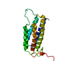
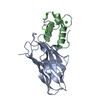
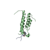
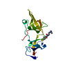
- PDB-1n7f: Crystal structure of the sixth PDZ domain of GRIP1 in complex wit... -
+ Open data

- Basic information
| Entry | Database: PDB / ID: 1n7f | ||||||
|---|---|---|---|---|---|---|---|
| Title | Crystal structure of the sixth PDZ domain of GRIP1 in complex with liprin C-terminal peptide | ||||||
 Components Components |
| ||||||
 Keywords Keywords | PROTEIN BINDING / PDZ / grip / liprin | ||||||
| Function / homology |  Function and homology information Function and homology informationregulation of synaptic scaling / exocyst / Receptor-type tyrosine-protein phosphatases / spine apparatus / Acetylcholine Neurotransmitter Release Cycle / Serotonin Neurotransmitter Release Cycle / vesicle-mediated transport in synapse / positive regulation of neuron projection arborization / neurotransmitter receptor transport, endosome to postsynaptic membrane / dendrite arborization ...regulation of synaptic scaling / exocyst / Receptor-type tyrosine-protein phosphatases / spine apparatus / Acetylcholine Neurotransmitter Release Cycle / Serotonin Neurotransmitter Release Cycle / vesicle-mediated transport in synapse / positive regulation of neuron projection arborization / neurotransmitter receptor transport, endosome to postsynaptic membrane / dendrite arborization / Norepinephrine Neurotransmitter Release Cycle / Dopamine Neurotransmitter Release Cycle / protein transporter activity / Glutamate Neurotransmitter Release Cycle / cortical microtubule organization / cellular response to phorbol 13-acetate 12-myristate / proximal dendrite / negative regulation of stress fiber assembly / positive regulation of dendrite morphogenesis / positive regulation of mini excitatory postsynaptic potential / long-term synaptic depression / presynaptic active zone / spine synapse / dendritic spine neck / dendritic spine head / Trafficking of GluR2-containing AMPA receptors / cellular response to glycine / negative regulation of protein localization to plasma membrane / regulation of postsynaptic membrane neurotransmitter receptor levels / synaptic cleft / cellular response to brain-derived neurotrophic factor stimulus / excitatory synapse / ionotropic glutamate receptor binding / cell-matrix adhesion / dendritic shaft / positive regulation of protein localization to plasma membrane / synaptic membrane / PDZ domain binding / cerebral cortex development / synapse organization / recycling endosome / Schaffer collateral - CA1 synapse / GABA-ergic synapse / terminal bouton / intracellular protein localization / long-term synaptic potentiation / nervous system development / presynapse / GTPase binding / presynaptic membrane / cell cortex / dendritic spine / microtubule / perikaryon / postsynaptic membrane / neuron projection / postsynaptic density / postsynapse / membrane raft / signaling receptor binding / axon / focal adhesion / neuronal cell body / dendrite / endoplasmic reticulum membrane / perinuclear region of cytoplasm / glutamatergic synapse / signal transduction / protein-containing complex / plasma membrane / cytoplasm / cytosol Similarity search - Function | ||||||
| Biological species |  | ||||||
| Method |  X-RAY DIFFRACTION / SYNCHROTRON / MOLECULAR REPLACEMENT / Resolution: 1.8 Å X-RAY DIFFRACTION / SYNCHROTRON / MOLECULAR REPLACEMENT / Resolution: 1.8 Å | ||||||
 Authors Authors | Im, Y.J. / Park, S.H. / Rho, S.H. / Lee, J.H. / Kang, G.B. / Sheng, M. / Kim, E. / Eom, S.H. | ||||||
 Citation Citation | Journal: J.BIOL.CHEM. / Year: 2003 Title: Crystal structure of GRIP1 PDZ6-peptide complex reveals the structural basis for class II PDZ target recognition and PDZ domain-mediated multimerization Authors: Im, Y.J. / Park, S.H. / Rho, S.H. / Lee, J.H. / Kang, G.B. / Sheng, M. / Kim, E. / Eom, S.H. | ||||||
| History |
|
- Structure visualization
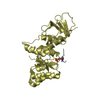
| Structure viewer | Molecule: MolmilJmol/JSmol |
|---|
- Downloads & links
-Download
| PDBx/mmCIF format | 1n7f.cif.gz | 52.4 KB | Display | PDBx/mmCIF format |
|---|---|---|---|---|
| PDB format | pdb1n7f.ent.gz | 38.5 KB | Display | PDB format |
| PDBx/mmJSON format | 1n7f.json.gz | Tree view | PDBx/mmJSON format | |
| Others |  Other downloads Other downloads |
-Validation report
| Arichive directory | https://data.pdbj.org/pub/pdb/validation_reports/n7/1n7fftp://data.pdbj.org/pub/pdb/validation_reports/n7/1n7f | HTTPS FTP |
|---|
-Related structure data


-Links
 PDBj
PDBj


- Assembly
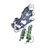
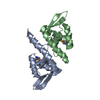
| Deposited unit | 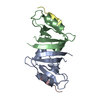
| ||||||||
|---|---|---|---|---|---|---|---|---|---|
| 1 |
| ||||||||
| Unit cell |
|
-Components
| #1: Protein | Mass: 10142.639 Da / Num. of mol.: 2 / Fragment: sixth PDZ domain Source method: isolated from a genetically manipulated source Source: (gene. exp.)  #2: Protein/peptide | Mass: 901.020 Da / Num. of mol.: 2 / Source method: obtained synthetically Details: The sequence of this chemically synthetized octa peptide occurs in the C-termiuns of human liprin alpha protein References: GenBank: 21707845, UniProt: Q13136*PLUS #3: Water | ChemComp-HOH / |  Mass: 18.015 Da / Num. of mol.: 236 / Source method: isolated from a natural source / Formula: H2O Mass: 18.015 Da / Num. of mol.: 236 / Source method: isolated from a natural source / Formula: H2O |
|---|
-Experimental details
-Experiment
| Experiment | Method: X-RAY DIFFRACTION / Number of used crystals: 1 |
|---|
- Sample preparation
| Crystal | Density Matthews: 2.96 Å3/Da / Density % sol: 58.09 % |
|---|---|
| Crystal grow | Temperature: 293 K / Method: vapor diffusion, hanging drop / pH: 5.5 Details: PEG400, MPD, pH 5.5, VAPOR DIFFUSION, HANGING DROP, temperature 293K |
| Crystal grow | *PLUS Details: Park, S.H., (2002) Acta Cryst., D58, 1063. |
-Data collection
| Diffraction | Mean temperature: 110 K |
|---|---|
| Diffraction source | Source: SYNCHROTRON / Site: Photon Factory  / Beamline: BL-18B / Wavelength: 1 Å / Beamline: BL-18B / Wavelength: 1 Å |
| Detector | Type: ADSC QUANTUM 4 / Detector: CCD / Date: Dec 13, 2000 |
| Radiation | Monochromator: Si 111 CHANNEL / Protocol: SINGLE WAVELENGTH / Monochromatic (M) / Laue (L): M / Scattering type: x-ray |
| Radiation wavelength | Wavelength: 1 Å / Relative weight: 1 |
| Reflection | Resolution: 1.8→50 Å / Num. all: 192446 / Num. obs: 191677 / % possible obs: 99.6 % / Observed criterion σ(F): 3 / Observed criterion σ(I): 3 / Redundancy: 7.58 % / Biso Wilson estimate: 17.5 Å2 / Rmerge(I) obs: 0.088 / Rsym value: 0.088 / Net I/σ(I): 10.1 |
| Reflection shell | Resolution: 1.8→1.89 Å / Rmerge(I) obs: 0.566 / Mean I/σ(I) obs: 4.5 / Rsym value: 0.566 / % possible all: 97.3 |
| Reflection shell | *PLUS % possible obs: 97.3 % |
- Processing
| Software |
| ||||||||||||||||||||||||||||||||||||||||||||||||||||||||||||
|---|---|---|---|---|---|---|---|---|---|---|---|---|---|---|---|---|---|---|---|---|---|---|---|---|---|---|---|---|---|---|---|---|---|---|---|---|---|---|---|---|---|---|---|---|---|---|---|---|---|---|---|---|---|---|---|---|---|---|---|---|---|
| Refinement | Method to determine structure: MOLECULAR REPLACEMENT / Resolution: 1.8→27.26 Å / Rfactor Rfree error: 0.005 / Isotropic thermal model: RESTRAINED / Cross valid method: THROUGHOUT / σ(F): 0 / σ(I): 0 / Stereochemistry target values: Engh & Huber Details: Structure solved by Br-MAD phasing of peptide free crystal
| ||||||||||||||||||||||||||||||||||||||||||||||||||||||||||||
| Solvent computation | Solvent model: FLAT MODEL / Bsol: 56.4386 Å2 / ksol: 0.35575 e/Å3 | ||||||||||||||||||||||||||||||||||||||||||||||||||||||||||||
| Displacement parameters | Biso mean: 29.4 Å2
| ||||||||||||||||||||||||||||||||||||||||||||||||||||||||||||
| Refine analyze |
| ||||||||||||||||||||||||||||||||||||||||||||||||||||||||||||
| Refinement step | Cycle: LAST / Resolution: 1.8→27.26 Å
| ||||||||||||||||||||||||||||||||||||||||||||||||||||||||||||
| Refine LS restraints |
| ||||||||||||||||||||||||||||||||||||||||||||||||||||||||||||
| LS refinement shell | Resolution: 1.8→1.91 Å / Total num. of bins used: 6
| ||||||||||||||||||||||||||||||||||||||||||||||||||||||||||||
| Xplor file |
| ||||||||||||||||||||||||||||||||||||||||||||||||||||||||||||
| Refinement | *PLUS Highest resolution: 1.8 Å / Lowest resolution: 15 Å / % reflection Rfree: 10 % / Rfactor Rwork: 0.201 | ||||||||||||||||||||||||||||||||||||||||||||||||||||||||||||
| Solvent computation | *PLUS | ||||||||||||||||||||||||||||||||||||||||||||||||||||||||||||
| Displacement parameters | *PLUS | ||||||||||||||||||||||||||||||||||||||||||||||||||||||||||||
| Refine LS restraints | *PLUS
|
