 Movie
Movie Controller

[English] 日本語
 Yorodumi
- PDB-2vky: Headbinding Domain of Phage P22 Tailspike C-Terminally Fused to I... -
+ Open data

- Basic information
| Entry | Database: PDB / ID: 2vky | ||||||
|---|---|---|---|---|---|---|---|
| Title | Headbinding Domain of Phage P22 Tailspike C-Terminally Fused to Isoleucine Zipper pIIGCN4 (Chimera I) | ||||||
 Components Components | TAIL PROTEIN, PIIGCN4 | ||||||
 Keywords Keywords | VIRAL PROTEIN / HEAD-BINDING DOMAIN / PHAGE P22 TAILSPIKE / CHIMERA / HYDROLASE / LATE PROTEIN / ISOLEUCINE ZIPPER PIIGCN4 | ||||||
| Function / homology |  Function and homology information Function and homology informationendo-1,3-alpha-L-rhamnosidase activity / symbiont entry into host cell via disruption of host cell envelope lipopolysaccharide / virus tail, fiber / symbiont entry into host cell via disruption of host cell envelope / symbiont entry into host / Hydrolases; Glycosylases; Glycosidases, i.e. enzymes that hydrolyse O- and S-glycosyl compounds / adhesion receptor-mediated virion attachment to host cell / virion attachment to host cell Similarity search - Function | ||||||
| Biological species |  ENTEROBACTERIA PHAGE P22 (virus) ENTEROBACTERIA PHAGE P22 (virus) | ||||||
| Method |  X-RAY DIFFRACTION / MOLECULAR REPLACEMENT / Resolution: 2.05 Å X-RAY DIFFRACTION / MOLECULAR REPLACEMENT / Resolution: 2.05 Å | ||||||
 Authors Authors | Seul, A. / Mueller, J.J. / Mueller, G. / Heinemann, U. / Seckler, R. | ||||||
 Citation Citation | Journal: Acta Crystallogr.,Sect.D / Year: 2014 Title: Bacteriophage P22 Tailspike: Structure of the Complete Protein and Function of the Interdomain Linker Authors: Seul, A. / Mueller, J.J. / Andres, D. / Stettner, E. / Heinemann, U. / Seckler, R. | ||||||
| History |
|
- Structure visualization
| Structure viewer | Molecule: MolmilJmol/JSmol |
|---|
- Downloads & links
-Download
| PDBx/mmCIF format | 2vky.cif.gz | 79.7 KB | Display | PDBx/mmCIF format |
|---|---|---|---|---|
| PDB format | pdb2vky.ent.gz | 60.4 KB | Display | PDB format |
| PDBx/mmJSON format | 2vky.json.gz | Tree view | PDBx/mmJSON format | |
| Others |  Other downloads Other downloads |
-Validation report
| Arichive directory | https://data.pdbj.org/pub/pdb/validation_reports/vk/2vkyftp://data.pdbj.org/pub/pdb/validation_reports/vk/2vky | HTTPS FTP |
|---|
-Related structure data
| Related structure data |  2vnlC  2xc1C  1eboS  1lktS S: Starting model for refinement C: citing same article ( |
|---|---|
| Similar structure data |


-Links
 PDBj
PDBj- Assembly
| Deposited unit | 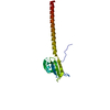
| |||||||||
|---|---|---|---|---|---|---|---|---|---|---|
| 1 | 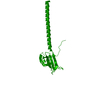
| |||||||||
| Unit cell |
| |||||||||
| Components on special symmetry positions |
|
-Components
| #1: Protein | Mass: 17216.551 Da / Num. of mol.: 1 / Fragment: HEAD-BINDING DOMAIN, RESIDUES 2-124 / Mutation: YES Source method: isolated from a genetically manipulated source Details: AMINO ACIDS 2-124 OF PHAGE P22 TAILSPIKE HAVE BEEN LINKED TO THE ISOLEUCINE ZIPPER PIIGCN4 125-155 Source: (gene. exp.) ENTEROBACTERIA PHAGE P22 (virus), (gene. exp.) Strain: SALMONELLA ENTERICA TYPHIMURIUM / Description: PIIGCN4, MODIFIED SEQUENCE NCBI GI\:5542583 / Plasmid: PET17B / Production host:  | ||
|---|---|---|---|
| #2: Water | ChemComp-HOH /  Mass: 18.015 Da / Num. of mol.: 173 / Source method: isolated from a natural source / Formula: H2O Mass: 18.015 Da / Num. of mol.: 173 / Source method: isolated from a natural source / Formula: H2O | ||
| Compound details | ENGINEERED| Sequence details | RESIDUES 125 TO 155 ARE NOT FOUND IN UNIPROT ENTRY P12528 BUT ARE PART OF GENBANK ENTRY GI:5542583. ...RESIDUES 125 TO 155 ARE NOT FOUND IN UNIPROT ENTRY P12528 BUT ARE PART OF GENBANK ENTRY GI:5542583. M1 OF GI5542583 IS MUTATED TO I125 IN THIS ENTRY. | |
-Experimental details
-Experiment
| Experiment | Method: X-RAY DIFFRACTION / Number of used crystals: 1 |
|---|
- Sample preparation
| Crystal | Density Matthews: 3 Å3/Da / Density % sol: 59 % / Description: NONE |
|---|---|
| Crystal grow | Method: vapor diffusion, hanging drop / pH: 6.5 Details: VAPOR DIFFUSION, HANGING DROP. PROTEIN: CONC. 8.2 MG/ML,BUFFER 50MM HEPES, PH6.5; RESERVOIR: 20% ISOPROPANOL, 0.1M NA-ACETATE, PH4.6, 0.2M CACL2; DROPLET 2 MICROL: 2 MICROL.CRYO:30% GLYCEROL. |
-Data collection
| Diffraction | Mean temperature: 110 K |
|---|---|
| Diffraction source | Source: ROTATING ANODE / Type: RIGAKU RU H2B / Wavelength: 1.5418 |
| Detector | Type: MARRESEARCH / Detector: IMAGE PLATE / Date: Sep 8, 2005 / Details: SLITS |
| Radiation | Monochromator: OSMIC MIRROR / Protocol: SINGLE WAVELENGTH / Monochromatic (M) / Laue (L): M / Scattering type: x-ray |
| Radiation wavelength | Wavelength: 1.5418 Å / Relative weight: 1 |
| Reflection | Resolution: 2.05→19.7 Å / Num. obs: 12716 / % possible obs: 99.6 % / Observed criterion σ(I): 0 / Redundancy: 4.2 % / Biso Wilson estimate: 30.86 Å2 / Rsym value: 0.03 / Net I/σ(I): 31.66 |
| Reflection shell | Resolution: 2.05→2.2 Å / Redundancy: 4.2 % / Mean I/σ(I) obs: 13.6 / Rsym value: 0.1 / % possible all: 99.5 |
- Processing
| Software |
| ||||||||||||||||||||||||||||||||||||||||||||||||||||||||||||||||||||||||||||||||||||||||||||||||||||||||||||||||||||||||||||||||||||||||||||||||||||||||||||||||||||||||||||||||||||||
|---|---|---|---|---|---|---|---|---|---|---|---|---|---|---|---|---|---|---|---|---|---|---|---|---|---|---|---|---|---|---|---|---|---|---|---|---|---|---|---|---|---|---|---|---|---|---|---|---|---|---|---|---|---|---|---|---|---|---|---|---|---|---|---|---|---|---|---|---|---|---|---|---|---|---|---|---|---|---|---|---|---|---|---|---|---|---|---|---|---|---|---|---|---|---|---|---|---|---|---|---|---|---|---|---|---|---|---|---|---|---|---|---|---|---|---|---|---|---|---|---|---|---|---|---|---|---|---|---|---|---|---|---|---|---|---|---|---|---|---|---|---|---|---|---|---|---|---|---|---|---|---|---|---|---|---|---|---|---|---|---|---|---|---|---|---|---|---|---|---|---|---|---|---|---|---|---|---|---|---|---|---|---|---|
| Refinement | Method to determine structure: MOLECULAR REPLACEMENT Starting model: PDB ENTRIES 1LKT, 1EBO Resolution: 2.05→20 Å / Cor.coef. Fo:Fc: 0.965 / Cor.coef. Fo:Fc free: 0.941 / SU B: 5.75 / SU ML: 0.083 / Cross valid method: THROUGHOUT / ESU R: 0.137 / ESU R Free: 0.131 / Stereochemistry target values: MAXIMUM LIKELIHOOD Details: HYDROGENS HAVE BEEN ADDED IN THE RIDING POSITIONS. THE HEAD-BINDING DOMAIN IS FUSED TO THE ISOLEUCINE ZIPPER PIIGCN4. THE BIOLOGICALLY ACTIVE HEAD- BINDING DOMAIN IS A CRYSTALLOGRAPHIC TRIMER.
| ||||||||||||||||||||||||||||||||||||||||||||||||||||||||||||||||||||||||||||||||||||||||||||||||||||||||||||||||||||||||||||||||||||||||||||||||||||||||||||||||||||||||||||||||||||||
| Solvent computation | Ion probe radii: 0.8 Å / Shrinkage radii: 0.8 Å / VDW probe radii: 1.2 Å / Solvent model: BABINET MODEL WITH MASK | ||||||||||||||||||||||||||||||||||||||||||||||||||||||||||||||||||||||||||||||||||||||||||||||||||||||||||||||||||||||||||||||||||||||||||||||||||||||||||||||||||||||||||||||||||||||
| Displacement parameters | Biso mean: 24.46 Å2
| ||||||||||||||||||||||||||||||||||||||||||||||||||||||||||||||||||||||||||||||||||||||||||||||||||||||||||||||||||||||||||||||||||||||||||||||||||||||||||||||||||||||||||||||||||||||
| Refinement step | Cycle: LAST / Resolution: 2.05→20 Å
| ||||||||||||||||||||||||||||||||||||||||||||||||||||||||||||||||||||||||||||||||||||||||||||||||||||||||||||||||||||||||||||||||||||||||||||||||||||||||||||||||||||||||||||||||||||||
| Refine LS restraints |
|