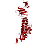
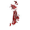
 Movie
Movie Controller


+ Open data

- Basic information
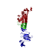
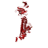
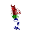
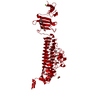
| Entry | Database: PDB / ID: 1qq1 | ||||||
|---|---|---|---|---|---|---|---|
| Title | TAILSPIKE PROTEIN, MUTANT E359G | ||||||
 Components Components | TAILSPIKE PROTEIN | ||||||
 Keywords Keywords | VIRAL PROTEIN / BETA-HELIX | ||||||
| Function / homology |  Function and homology information Function and homology informationendo-1,3-alpha-L-rhamnosidase activity / symbiont entry into host cell via disruption of host cell envelope lipopolysaccharide / virus tail, fiber / symbiont entry into host cell via disruption of host cell envelope / symbiont entry into host / Hydrolases; Glycosylases; Glycosidases, i.e. enzymes that hydrolyse O- and S-glycosyl compounds / adhesion receptor-mediated virion attachment to host cell / virion attachment to host cell Similarity search - Function | ||||||
| Biological species |  Enterobacteria phage P22 (virus) Enterobacteria phage P22 (virus) | ||||||
| Method |  X-RAY DIFFRACTION / Resolution: 1.8 Å X-RAY DIFFRACTION / Resolution: 1.8 Å | ||||||
 Authors Authors | Schuler, B. / Furst, F. / Osterroth, F. / Steinbacher, S. / Huber, R. / Seckler, R. | ||||||
 Citation Citation | Journal: Proteins / Year: 2000 Title: Plasticity and steric strain in a parallel beta-helix: rational mutations in the P22 tailspike protein. Authors: Schuler, B. / Furst, F. / Osterroth, F. / Steinbacher, S. / Huber, R. / Seckler, R. | ||||||
| History |
|
- Structure visualization
| Structure viewer | Molecule: MolmilJmol/JSmol |
|---|
- Downloads & links
-Download
| PDBx/mmCIF format | 1qq1.cif.gz | 118 KB | Display | PDBx/mmCIF format |
|---|---|---|---|---|
| PDB format | pdb1qq1.ent.gz | 89.8 KB | Display | PDB format |
| PDBx/mmJSON format | 1qq1.json.gz | Tree view | PDBx/mmJSON format | |
| Others |  Other downloads Other downloads |
-Validation report
| Arichive directory | https://data.pdbj.org/pub/pdb/validation_reports/qq/1qq1ftp://data.pdbj.org/pub/pdb/validation_reports/qq/1qq1 | HTTPS FTP |
|---|
-Related structure data


-Links
 PDBj
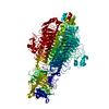
PDBj- Assembly
| Deposited unit | 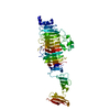
| ||||||||
|---|---|---|---|---|---|---|---|---|---|
| 1 | 
| ||||||||
| Unit cell |
|
-Components
| #1: Protein | Mass: 60193.207 Da / Num. of mol.: 1 / Fragment: C-TERMINAL FRAGMENT / Mutation: E359G Source method: isolated from a genetically manipulated source Source: (gene. exp.) Enterobacteria phage P22 (virus) / Genus: P22-like viruses / References: UniProt: P12528 |
|---|---|
| #2: Water | ChemComp-HOH /  Mass: 18.015 Da / Num. of mol.: 217 / Source method: isolated from a natural source / Formula: H2O Mass: 18.015 Da / Num. of mol.: 217 / Source method: isolated from a natural source / Formula: H2O |
-Experimental details
-Experiment
| Experiment | Method: X-RAY DIFFRACTION / Number of used crystals: 1 |
|---|
- Sample preparation
| Crystal | Density Matthews: 2.45 Å3/Da / Density % sol: 49.71 % | |||||||||||||||||||||||||||||||||||
|---|---|---|---|---|---|---|---|---|---|---|---|---|---|---|---|---|---|---|---|---|---|---|---|---|---|---|---|---|---|---|---|---|---|---|---|---|
| Crystal grow | Temperature: 293 K / Method: vapor diffusion, hanging drop / pH: 10 Details: 1M AMMONIUM SULPHATE, 0.1M SODIUM PHOSPHATE, pH 10.0, VAPOR DIFFUSION, HANGING DROP, temperature 293K | |||||||||||||||||||||||||||||||||||
| Crystal grow | *PLUS Temperature: 4 ℃ / Details: Steinbacher, S., (1994) Science, 265, 383. | |||||||||||||||||||||||||||||||||||
| Components of the solutions | *PLUS
|
-Data collection
| Diffraction | Mean temperature: 287 K |
|---|---|
| Diffraction source | Source: ROTATING ANODE / Type: RIGAKU RU200 / Wavelength: 1.54 |
| Detector | Type: MARRESEARCH / Detector: AREA DETECTOR / Date: Aug 12, 1998 |
| Radiation | Protocol: SINGLE WAVELENGTH / Monochromatic (M) / Laue (L): M / Scattering type: x-ray |
| Radiation wavelength | Wavelength: 1.54 Å / Relative weight: 1 |
| Reflection | Resolution: 1.8→8 Å / Num. all: 53301 / Num. obs: 53301 / % possible obs: 98.8 % / Observed criterion σ(F): 0 / Observed criterion σ(I): 0 / Redundancy: 4.3 % / Biso Wilson estimate: 20.4 Å2 / Rmerge(I) obs: 0.071 / Net I/σ(I): 10.3 |
| Reflection shell | Resolution: 1.8→1.84 Å / Redundancy: 4.5 % / Rmerge(I) obs: 0.324 / % possible all: 98.3 |
| Reflection | *PLUS Num. measured all: 232408 |
| Reflection shell | *PLUS % possible obs: 98.3 % |
- Processing
| Software |
| ||||||||||||||||||||||||||||||||||||||||||||||||||||||||||||
|---|---|---|---|---|---|---|---|---|---|---|---|---|---|---|---|---|---|---|---|---|---|---|---|---|---|---|---|---|---|---|---|---|---|---|---|---|---|---|---|---|---|---|---|---|---|---|---|---|---|---|---|---|---|---|---|---|---|---|---|---|---|
| Refinement | Resolution: 1.8→8 Å / σ(F): 0 / σ(I): 0 / Stereochemistry target values: ENGH & HUBER
| ||||||||||||||||||||||||||||||||||||||||||||||||||||||||||||
| Refinement step | Cycle: LAST / Resolution: 1.8→8 Å
| ||||||||||||||||||||||||||||||||||||||||||||||||||||||||||||
| Refine LS restraints |
| ||||||||||||||||||||||||||||||||||||||||||||||||||||||||||||
| Software | *PLUS Name: X-PLOR / Version: 3.851 / Classification: refinement | ||||||||||||||||||||||||||||||||||||||||||||||||||||||||||||
| Refinement | *PLUS Lowest resolution: 8 Å / σ(F): 0 | ||||||||||||||||||||||||||||||||||||||||||||||||||||||||||||
| Solvent computation | *PLUS | ||||||||||||||||||||||||||||||||||||||||||||||||||||||||||||
| Displacement parameters | *PLUS | ||||||||||||||||||||||||||||||||||||||||||||||||||||||||||||
| Refine LS restraints | *PLUS
|
