 Movie
Movie Controller

[English] 日本語
 Yorodumi
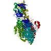
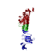
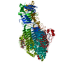
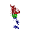
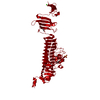
- PDB-2vfn: Low Temperature Structure of P22 Tailspike Protein Fragment (109-... -
+ Open data

- Basic information
| Entry | Database: PDB / ID: 2vfn | ||||||
|---|---|---|---|---|---|---|---|
| Title | Low Temperature Structure of P22 Tailspike Protein Fragment (109-666), Mutant V125A | ||||||
 Components Components | BIFUNCTIONAL TAIL PROTEIN | ||||||
 Keywords Keywords | HYDROLASE / P22 TAILSPIKE PROTEIN / SALMONELLA BACTERIOPHAGE P22 / PROTEIN FOLDING / PROTEIN STABILITY / RIGHT-HANDED PARALLEL BETA-HELIX / LATE PROTEIN / ENDOGLYCOSIDASE | ||||||
| Function / homology |  Function and homology information Function and homology informationendo-1,3-alpha-L-rhamnosidase activity / symbiont entry into host cell via disruption of host cell envelope lipopolysaccharide / virus tail, fiber / symbiont entry into host cell via disruption of host cell envelope / symbiont entry into host / Hydrolases; Glycosylases; Glycosidases, i.e. enzymes that hydrolyse O- and S-glycosyl compounds / adhesion receptor-mediated virion attachment to host cell / virion attachment to host cell Similarity search - Function | ||||||
| Biological species |  ENTEROBACTERIA PHAGE P22 (virus) ENTEROBACTERIA PHAGE P22 (virus) | ||||||
| Method |  X-RAY DIFFRACTION / SYNCHROTRON / MOLECULAR REPLACEMENT / Resolution: 1.5 Å X-RAY DIFFRACTION / SYNCHROTRON / MOLECULAR REPLACEMENT / Resolution: 1.5 Å | ||||||
 Authors Authors | Becker, M. / Mueller, J.J. / Heinemann, U. / Seckler, R. | ||||||
 Citation Citation | Journal: To be Published Title: Side-Chain Stacking and Beta-Helix Stability in P22 Tailspike Protein Authors: Becker, M. / Mueller, J.J. / Weikl, T. / Heinemann, U. / Seckler, R. #1: Journal: Proteins: Struct.,Funct., Genet. / Year: 2000Title: Plasticity and Steric Strain in a Parallel Beta-Helix: Rational Mutations in the P22 Tailspike Protein Authors: Schuler, B. / Fuerst, F. / Osterroth, F. / Steinbacher, S. / Huber, R. / Seckler, R. | ||||||
| History |
| ||||||
| Remark 650 | HELIX DETERMINATION METHOD: AUTHOR PROVIDED. | ||||||
| Remark 700 | SHEET DETERMINATION METHOD: AUTHOR PROVIDED. |
- Structure visualization
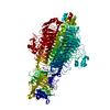
| Structure viewer | Molecule: MolmilJmol/JSmol |
|---|
- Downloads & links
-Download
| PDBx/mmCIF format | 2vfn.cif.gz | 145.2 KB | Display | PDBx/mmCIF format |
|---|---|---|---|---|
| PDB format | pdb2vfn.ent.gz | 111.5 KB | Display | PDB format |
| PDBx/mmJSON format | 2vfn.json.gz | Tree view | PDBx/mmJSON format | |
| Others |  Other downloads Other downloads |
-Validation report
| Arichive directory | https://data.pdbj.org/pub/pdb/validation_reports/vf/2vfnftp://data.pdbj.org/pub/pdb/validation_reports/vf/2vfn | HTTPS FTP |
|---|
-Related structure data
| Related structure data |  2vfmC  2vfoC  2vfpC  2vfqSC C: citing same article ( S: Starting model for refinement |
|---|---|
| Similar structure data |


-Links
 PDBj
PDBj- Assembly
| Deposited unit | 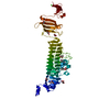
| ||||||||||||||||||||||||
|---|---|---|---|---|---|---|---|---|---|---|---|---|---|---|---|---|---|---|---|---|---|---|---|---|---|
| 1 | 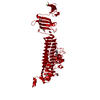
| ||||||||||||||||||||||||
| Unit cell |
| ||||||||||||||||||||||||
| Components on special symmetry positions |
|
-Components
| #1: Protein | Mass: 60205.238 Da / Num. of mol.: 1 / Fragment: RESIDUES 110-667 / Mutation: YES Source method: isolated from a genetically manipulated source Details: THE PROTEIN IS LACKING THE N-TERMINAL HEAD-BINDING DOMAIN Source: (gene. exp.) ENTEROBACTERIA PHAGE P22 (virus) / Plasmid: P125ADN / Production host:  References: UniProt: P12528, Hydrolases; Glycosylases; Glycosidases, i.e. enzymes that hydrolyse O- and S-glycosyl compounds | ||||||||||
|---|---|---|---|---|---|---|---|---|---|---|---|
| #2: Chemical | ChemComp-GOL /   Mass: 92.094 Da / Num. of mol.: 10 / Source method: obtained synthetically / Formula: C3H8O3 Mass: 92.094 Da / Num. of mol.: 10 / Source method: obtained synthetically / Formula: C3H8O3#3: Chemical | ChemComp-SO4 / |   Mass: 96.063 Da / Num. of mol.: 1 / Source method: obtained synthetically / Formula: SO4 Mass: 96.063 Da / Num. of mol.: 1 / Source method: obtained synthetically / Formula: SO4#4: Chemical | ChemComp-CA / |   Mass: 40.078 Da / Num. of mol.: 1 / Source method: obtained synthetically / Formula: Ca Mass: 40.078 Da / Num. of mol.: 1 / Source method: obtained synthetically / Formula: Ca#5: Water | ChemComp-HOH / |  Mass: 18.015 Da / Num. of mol.: 804 / Source method: isolated from a natural source / Formula: H2O Mass: 18.015 Da / Num. of mol.: 804 / Source method: isolated from a natural source / Formula: H2OCompound details | ENGINEERED | Sequence details | GLY513 IN THE SWISSPROT ENTRY WAS CORRECTED IN THE SEQUENCE TO SER513 WHICH IS ALSO PRESENT IN THE ...GLY513 IN THE SWISSPROT ENTRY WAS CORRECTED IN THE SEQUENCE TO SER513 WHICH IS ALSO PRESENT IN THE PARENT WILD-TYPE DNA | |
-Experimental details
-Experiment
| Experiment | Method: X-RAY DIFFRACTION / Number of used crystals: 1 |
|---|
- Sample preparation
| Crystal | Density Matthews: 2.44 Å3/Da / Density % sol: 49.49 % Description: FOR REFINEMENT REFLECTIONS IN THE CORNERS OF THE IMAGE PLATE WERE USED ADDITIONALLY, 1.5-1.59 A RESOLUTION, COMPETENESS 42.6 PERCENT, REDUNDANCY 1.7, RSYM 0.101, I DIVIDED BY SIGMA(I) 6.8 |
|---|---|
| Crystal grow | pH: 10 Details: DROP: 2 MICROLITER 1.5 M AMMONIUM SULFATE, 0.1 M SODIUM PHOSPHATE, PH 10.0, PLUS 3.3 MICROLITER, 10 MG/ML PROTEIN SOLUTION IN 10 MM HEPES, PH 7.0; RESERVOIR: 750 MICOLITER 1.0 M AMMONIUM ...Details: DROP: 2 MICROLITER 1.5 M AMMONIUM SULFATE, 0.1 M SODIUM PHOSPHATE, PH 10.0, PLUS 3.3 MICROLITER, 10 MG/ML PROTEIN SOLUTION IN 10 MM HEPES, PH 7.0; RESERVOIR: 750 MICOLITER 1.0 M AMMONIUM SULFATE, 0.1 M SODIUM PHOSPHATE, PH 10.0 |
-Data collection
| Diffraction | Mean temperature: 100 K |
|---|---|
| Diffraction source | Source: SYNCHROTRON / Site: BESSY  / Beamline: 14.1 / Wavelength: 0.92018 / Beamline: 14.1 / Wavelength: 0.92018 |
| Detector | Type: MARRESEARCH / Detector: CCD / Date: May 2, 2007 / Details: MIRRORS |
| Radiation | Monochromator: SI-111 / Protocol: SINGLE WAVELENGTH / Monochromatic (M) / Laue (L): M / Scattering type: x-ray |
| Radiation wavelength | Wavelength: 0.92018 Å / Relative weight: 1 |
| Reflection | Resolution: 1.59→50 Å / Num. obs: 76620 / % possible obs: 98.1 % / Redundancy: 12.9 % / Biso Wilson estimate: 18.4 Å2 / Rmerge(I) obs: 0.05 / Net I/σ(I): 34.9 |
| Reflection shell | Resolution: 1.59→1.7 Å / Redundancy: 3.6 % / Rmerge(I) obs: 0.1 / Mean I/σ(I) obs: 10.2 / % possible all: 90.5 |
- Processing
| Software |
| ||||||||||||||||||||||||||||||||||||||||||||||||||||||||||||||||||||||||||||||||||||||||||||||||||||||||||||||||||||||||||||||||||||||||||||||||||||||||||||||||||||||||||||||||||||||
|---|---|---|---|---|---|---|---|---|---|---|---|---|---|---|---|---|---|---|---|---|---|---|---|---|---|---|---|---|---|---|---|---|---|---|---|---|---|---|---|---|---|---|---|---|---|---|---|---|---|---|---|---|---|---|---|---|---|---|---|---|---|---|---|---|---|---|---|---|---|---|---|---|---|---|---|---|---|---|---|---|---|---|---|---|---|---|---|---|---|---|---|---|---|---|---|---|---|---|---|---|---|---|---|---|---|---|---|---|---|---|---|---|---|---|---|---|---|---|---|---|---|---|---|---|---|---|---|---|---|---|---|---|---|---|---|---|---|---|---|---|---|---|---|---|---|---|---|---|---|---|---|---|---|---|---|---|---|---|---|---|---|---|---|---|---|---|---|---|---|---|---|---|---|---|---|---|---|---|---|---|---|---|---|
| Refinement | Method to determine structure: MOLECULAR REPLACEMENT Starting model: PDB ENTRY 2VFQ Resolution: 1.5→49.15 Å / Cor.coef. Fo:Fc: 0.977 / Cor.coef. Fo:Fc free: 0.965 / SU B: 0.946 / SU ML: 0.036 / Cross valid method: THROUGHOUT / ESU R: 0.058 / ESU R Free: 0.061 / Stereochemistry target values: MAXIMUM LIKELIHOOD / Details: HYDROGENS HAVE BEEN ADDED IN THE RIDING POSITIONS.
| ||||||||||||||||||||||||||||||||||||||||||||||||||||||||||||||||||||||||||||||||||||||||||||||||||||||||||||||||||||||||||||||||||||||||||||||||||||||||||||||||||||||||||||||||||||||
| Solvent computation | Ion probe radii: 0.8 Å / Shrinkage radii: 0.8 Å / VDW probe radii: 1.4 Å / Solvent model: BABINET MODEL WITH MASK | ||||||||||||||||||||||||||||||||||||||||||||||||||||||||||||||||||||||||||||||||||||||||||||||||||||||||||||||||||||||||||||||||||||||||||||||||||||||||||||||||||||||||||||||||||||||
| Displacement parameters | Biso mean: 10.84 Å2 | ||||||||||||||||||||||||||||||||||||||||||||||||||||||||||||||||||||||||||||||||||||||||||||||||||||||||||||||||||||||||||||||||||||||||||||||||||||||||||||||||||||||||||||||||||||||
| Refinement step | Cycle: LAST / Resolution: 1.5→49.15 Å
| ||||||||||||||||||||||||||||||||||||||||||||||||||||||||||||||||||||||||||||||||||||||||||||||||||||||||||||||||||||||||||||||||||||||||||||||||||||||||||||||||||||||||||||||||||||||
| Refine LS restraints |
|