 Movie
Movie Controller

[English] 日本語
 Yorodumi
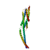
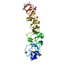
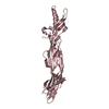
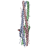
- PDB-1ebo: CRYSTAL STRUCTURE OF THE EBOLA VIRUS MEMBRANE-FUSION SUBUNIT, GP2... -
+ Open data

- Basic information
| Entry | Database: PDB / ID: 1ebo | ||||||
|---|---|---|---|---|---|---|---|
| Title | CRYSTAL STRUCTURE OF THE EBOLA VIRUS MEMBRANE-FUSION SUBUNIT, GP2, FROM THE ENVELOPE GLYCOPROTEIN ECTODOMAIN | ||||||
 Components Components | EBOLA VIRUS ENVELOPE PROTEIN CHIMERA CONSISTING OF A FRAGMENT OF GCN4 ZIPPER CLONED N-TERMINAL TO A FRAGMENT OF GP2 | ||||||
 Keywords Keywords | VIRAL PROTEIN / MEMBRANE FUSION SUBUNIT | ||||||
| Function / homology |  Function and homology information Function and homology informationsymbiont-mediated-mediated suppression of host tetherin activity / clathrin-dependent endocytosis of virus by host cell / entry receptor-mediated virion attachment to host cell / symbiont-mediated suppression of host innate immune response / fusion of virus membrane with host endosome membrane / viral envelope / lipid binding / host cell plasma membrane / virion membrane / extracellular region Similarity search - Function | ||||||
| Biological species |  Ebola virus sp. Ebola virus sp. | ||||||
| Method |  X-RAY DIFFRACTION / SIR / Resolution: 3 Å X-RAY DIFFRACTION / SIR / Resolution: 3 Å | ||||||
 Authors Authors | Weissenhorn, W. / Carfi, A. / Lee, K.H. / Skehel, J.J. / Wiley, D.C. | ||||||
 Citation Citation | Journal: Mol.Cell / Year: 1998 Title: Crystal structure of the Ebola virus membrane fusion subunit, GP2, from the envelope glycoprotein ectodomain. Authors: Weissenhorn, W. / Carfi, A. / Lee, K.H. / Skehel, J.J. / Wiley, D.C. | ||||||
| History |
|
- Structure visualization
| Structure viewer | Molecule: MolmilJmol/JSmol |
|---|
- Downloads & links
-Download
| PDBx/mmCIF format | 1ebo.cif.gz | 130.6 KB | Display | PDBx/mmCIF format |
|---|---|---|---|---|
| PDB format | pdb1ebo.ent.gz | 104.8 KB | Display | PDB format |
| PDBx/mmJSON format | 1ebo.json.gz | Tree view | PDBx/mmJSON format | |
| Others |  Other downloads Other downloads |
-Validation report
| Arichive directory | https://data.pdbj.org/pub/pdb/validation_reports/eb/1eboftp://data.pdbj.org/pub/pdb/validation_reports/eb/1ebo | HTTPS FTP |
|---|
-Related structure data
| Similar structure data |
|---|


-Links
 PDBj
PDBj

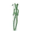
- Assembly
| Deposited unit | 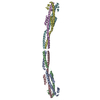
| ||||||||||
|---|---|---|---|---|---|---|---|---|---|---|---|
| 1 | 
| ||||||||||
| 2 | 
| ||||||||||
| 3 | 
| ||||||||||
| Unit cell |
|
-Components
| #1: Protein | Mass: 15359.446 Da / Num. of mol.: 6 / Fragment: GCN4 IS RESIDUE 3 - 32, GP2 IS RESIDUE 51 - 133 / Mutation: C55E, C108R Source method: isolated from a genetically manipulated source Source: (gene. exp.) Ebola virus sp. / Genus: Ebola-like viruses / Production host:  References: UniProt: Q913A3, GenBank: 4389202, GenBank: 4389203, GenBank: 4389204, GenBank: 4389205, GenBank: 4389206, UniProt: O11457*PLUS #2: Chemical |   Mass: 65.409 Da / Num. of mol.: 3 / Source method: obtained synthetically / Formula: Zn Mass: 65.409 Da / Num. of mol.: 3 / Source method: obtained synthetically / Formula: Zn#3: Chemical |   Mass: 35.453 Da / Num. of mol.: 2 / Source method: obtained synthetically / Formula: Cl Mass: 35.453 Da / Num. of mol.: 2 / Source method: obtained synthetically / Formula: ClHas protein modification | Y | |
|---|
-Experimental details
-Experiment
| Experiment | Method: X-RAY DIFFRACTION / Number of used crystals: 1 |
|---|
- Sample preparation
| Crystal | Density Matthews: 2.24 Å3/Da / Density % sol: 35 % | |||||||||||||||||||||||||||||||||||
|---|---|---|---|---|---|---|---|---|---|---|---|---|---|---|---|---|---|---|---|---|---|---|---|---|---|---|---|---|---|---|---|---|---|---|---|---|
| Crystal grow | pH: 8.5 / Details: pH 8.5 | |||||||||||||||||||||||||||||||||||
| Crystal grow | *PLUS pH: 9.3 / Method: vapor diffusion, hanging dropDetails: 0.02ml of protein solution was mixed with 0.01ml of reservoir solution | |||||||||||||||||||||||||||||||||||
| Components of the solutions | *PLUS
|
-Data collection
| Diffraction | Mean temperature: 290 K |
|---|---|
| Diffraction source | Source: ROTATING ANODE / Type: ELLIOTT GX-13 / Wavelength: 1.5418 |
| Detector | Type: MARRESEARCH / Detector: IMAGE PLATE / Date: Aug 15, 1998 / Details: MIRRORS |
| Radiation | Protocol: SINGLE WAVELENGTH / Monochromatic (M) / Laue (L): M / Scattering type: x-ray |
| Radiation wavelength | Wavelength: 1.5418 Å / Relative weight: 1 |
| Reflection | Resolution: 3→20 Å / Num. obs: 17123 / % possible obs: 99.3 % / Observed criterion σ(I): -3 / Redundancy: 5.1 % / Rsym value: 0.083 |
| Reflection shell | Resolution: 3→3.09 Å / Redundancy: 3.07 % / Rsym value: 0.29 / % possible all: 99.2 |
| Reflection | *PLUS Num. measured all: 87380 / Rmerge(I) obs: 0.083 |
| Reflection shell | *PLUS % possible obs: 99.2 % / Rmerge(I) obs: 0.29 |
- Processing
| Software |
| ||||||||||||||||||||||||||||||||||||||||||||||||||||||||||||
|---|---|---|---|---|---|---|---|---|---|---|---|---|---|---|---|---|---|---|---|---|---|---|---|---|---|---|---|---|---|---|---|---|---|---|---|---|---|---|---|---|---|---|---|---|---|---|---|---|---|---|---|---|---|---|---|---|---|---|---|---|---|
| Refinement | Method to determine structure: SIR / Resolution: 3→20 Å / Cross valid method: THROUGHOUT / σ(F): 2 Details: REFINEMENT WAS CONCLUDED USING CNS. A BULK SOLVENT CORRECTION WAS APPLIED
| ||||||||||||||||||||||||||||||||||||||||||||||||||||||||||||
| Refinement step | Cycle: LAST / Resolution: 3→20 Å
| ||||||||||||||||||||||||||||||||||||||||||||||||||||||||||||
| Refine LS restraints |
| ||||||||||||||||||||||||||||||||||||||||||||||||||||||||||||
| Software | *PLUS Name: X-PLOR / Version: 3.851 / Classification: refinement | ||||||||||||||||||||||||||||||||||||||||||||||||||||||||||||
| Refinement | *PLUS | ||||||||||||||||||||||||||||||||||||||||||||||||||||||||||||
| Solvent computation | *PLUS | ||||||||||||||||||||||||||||||||||||||||||||||||||||||||||||
| Displacement parameters | *PLUS Biso mean: 38.7 Å2 |
