 Movie
Movie Controller

[English] 日本語
 Yorodumi
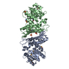
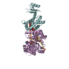
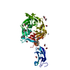
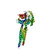
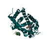
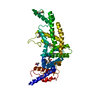
- PDB-6tix: Crystal structure of penicillin-binding protein 2 from Yersinia p... -
+ Open data

- Basic information
| Entry | Database: PDB / ID: 6tix | ||||||
|---|---|---|---|---|---|---|---|
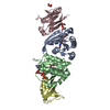
| Title | Crystal structure of penicillin-binding protein 2 from Yersinia pestis in complex with mecillinam | ||||||
 Components Components | Peptidoglycan D,D-transpeptidase MrdA | ||||||
 Keywords Keywords | MEMBRANE PROTEIN / Class B PBP / Yersinia pestis / HMM transpeptidase / periplasmic protein / peptidoglycan crosslinking / mecillinam | ||||||
| Function / homology |  Function and homology information Function and homology informationpeptidoglycan L,D-transpeptidase activity / serine-type D-Ala-D-Ala carboxypeptidase / serine-type D-Ala-D-Ala carboxypeptidase activity / penicillin binding / peptidoglycan biosynthetic process / cell wall organization / regulation of cell shape / proteolysis / plasma membrane Similarity search - Function | ||||||
| Biological species |   Yersinia pestis (bacteria) Yersinia pestis (bacteria) | ||||||
| Method |  X-RAY DIFFRACTION / SYNCHROTRON / MOLECULAR REPLACEMENT / Resolution: 2.8 Å X-RAY DIFFRACTION / SYNCHROTRON / MOLECULAR REPLACEMENT / Resolution: 2.8 Å | ||||||
 Authors Authors | Pankov, G. / Hunter, W.N. / Dawson, A. | ||||||
 Citation Citation | Journal: To Be Published Title: The structure of penicillin-binding protein 2 from Yersinia pestis in complex with mecillinam Authors: Pankov, G. | ||||||
| History |
|
- Structure visualization
| Structure viewer | Molecule: MolmilJmol/JSmol |
|---|
- Downloads & links
-Download
| PDBx/mmCIF format | 6tix.cif.gz | 223.7 KB | Display | PDBx/mmCIF format |
|---|---|---|---|---|
| PDB format | pdb6tix.ent.gz | Display | PDB format | |
| PDBx/mmJSON format | 6tix.json.gz | Tree view | PDBx/mmJSON format | |
| Others |  Other downloads Other downloads |
-Validation report
| Arichive directory | https://data.pdbj.org/pub/pdb/validation_reports/ti/6tixftp://data.pdbj.org/pub/pdb/validation_reports/ti/6tix | HTTPS FTP |
|---|
-Related structure data
| Related structure data | 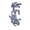 5lp4S S: Starting model for refinement |
|---|---|
| Similar structure data |


-Links
 PDBj
PDBj
- Assembly
| Deposited unit | 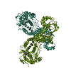
| ||||||||
|---|---|---|---|---|---|---|---|---|---|
| 1 | 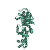
| ||||||||
| 2 | 
| ||||||||
| Unit cell |
| ||||||||
| Noncrystallographic symmetry (NCS) | NCS domain: (Details: Chains AAA BBB) |
-Components
| #1: Protein | Mass: 65552.883 Da / Num. of mol.: 2 Source method: isolated from a genetically manipulated source Source: (gene. exp.) Yersinia pestis (bacteria) / Gene: mrdA, YPO2604 / Production host: References: UniProt: A0A384KFW3, UniProt: A0A5P8YJF3*PLUS, serine-type D-Ala-D-Ala carboxypeptidase #2: Chemical | 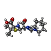  Mass: 327.442 Da / Num. of mol.: 2 / Source method: obtained synthetically / Formula: C15H25N3O3S Mass: 327.442 Da / Num. of mol.: 2 / Source method: obtained synthetically / Formula: C15H25N3O3S#3: Water | ChemComp-HOH / |  Mass: 18.015 Da / Num. of mol.: 104 / Source method: isolated from a natural source / Formula: H2O Mass: 18.015 Da / Num. of mol.: 104 / Source method: isolated from a natural source / Formula: H2OHas ligand of interest | Y | Has protein modification | Y | |
|---|
-Experimental details
-Experiment
| Experiment | Method: X-RAY DIFFRACTION / Number of used crystals: 1 |
|---|
- Sample preparation
| Crystal | Density Matthews: 2.35 Å3/Da / Density % sol: 47.6 % / Description: Branched rectangular prisms |
|---|---|
| Crystal grow | Temperature: 296 K / Method: vapor diffusion, hanging drop Details: Crystals were grown in a condition containing 1 uL of 0.8 mg/mL YpPBP2 (in 20 mM Tris-HCl pH 7.5, 150 mM NaCl, 2 mM mecillinam) and 1 uL of reservoir solution (0.45 M Na+/K+ tartrate, 0.1 M ...Details: Crystals were grown in a condition containing 1 uL of 0.8 mg/mL YpPBP2 (in 20 mM Tris-HCl pH 7.5, 150 mM NaCl, 2 mM mecillinam) and 1 uL of reservoir solution (0.45 M Na+/K+ tartrate, 0.1 M HEPES-NaOH pH 7.5, 7.2% PEG 4000). The size of these crystals was optimised by three rounds of macroseeding into condition containing (0.45 M Na+/K+ tartrate, 0.1 M HEPES-NaOH pH 7.5, 7.2% PEG 4000 and 2 mM mecillinam) |
-Data collection
| Diffraction | Mean temperature: 100 K / Serial crystal experiment: N |
|---|---|
| Diffraction source | Source: SYNCHROTRON / Site: ESRF  / Beamline: ID23-2 / Wavelength: 0.8731 Å / Beamline: ID23-2 / Wavelength: 0.8731 Å |
| Detector | Type: DECTRIS PILATUS3 X 2M / Detector: PIXEL / Date: Apr 13, 2018 |
| Radiation | Protocol: SINGLE WAVELENGTH / Monochromatic (M) / Laue (L): M / Scattering type: x-ray |
| Radiation wavelength | Wavelength: 0.8731 Å / Relative weight: 1 |
| Reflection | Resolution: 2.8→45.693 Å / Num. obs: 27215 / % possible obs: 92.9 % / Redundancy: 2.1 % / CC1/2: 0.991 / Rmerge(I) obs: 0.108 / Rpim(I) all: 0.097 / Net I/σ(I): 4.1 |
| Reflection shell | Resolution: 2.8→2.95 Å / Redundancy: 2.1 % / Rmerge(I) obs: 0.452 / Mean I/σ(I) obs: 1.2 / Num. unique obs: 4118 / CC1/2: 0.677 / Rpim(I) all: 0.452 / Rrim(I) all: 0.64 / % possible all: 96.7 |
- Processing
| Software |
| |||||||||||||||||||||||||||||||||||||||||||||||||||||||||||||||||||||||||||||||||||||||||||||||||||||||||||||||||||||||||||||||||||||||||||||||||||||||||||||||||||||
|---|---|---|---|---|---|---|---|---|---|---|---|---|---|---|---|---|---|---|---|---|---|---|---|---|---|---|---|---|---|---|---|---|---|---|---|---|---|---|---|---|---|---|---|---|---|---|---|---|---|---|---|---|---|---|---|---|---|---|---|---|---|---|---|---|---|---|---|---|---|---|---|---|---|---|---|---|---|---|---|---|---|---|---|---|---|---|---|---|---|---|---|---|---|---|---|---|---|---|---|---|---|---|---|---|---|---|---|---|---|---|---|---|---|---|---|---|---|---|---|---|---|---|---|---|---|---|---|---|---|---|---|---|---|---|---|---|---|---|---|---|---|---|---|---|---|---|---|---|---|---|---|---|---|---|---|---|---|---|---|---|---|---|---|---|---|---|
| Refinement | Method to determine structure: MOLECULAR REPLACEMENT Starting model: 5LP4 Resolution: 2.8→45.693 Å / Cor.coef. Fo:Fc: 0.938 / Cor.coef. Fo:Fc free: 0.891 / WRfactor Rfree: 0.274 / WRfactor Rwork: 0.214 / SU B: 29.964 / SU ML: 0.529 / Average fsc free: 0.7294 / Average fsc work: 0.772 / Cross valid method: FREE R-VALUE / ESU R Free: 0.48 Details: Hydrogens have been added in their riding positions
| |||||||||||||||||||||||||||||||||||||||||||||||||||||||||||||||||||||||||||||||||||||||||||||||||||||||||||||||||||||||||||||||||||||||||||||||||||||||||||||||||||||
| Solvent computation | Ion probe radii: 0.8 Å / Shrinkage radii: 0.8 Å / VDW probe radii: 1.2 Å / Solvent model: MASK BULK SOLVENT | |||||||||||||||||||||||||||||||||||||||||||||||||||||||||||||||||||||||||||||||||||||||||||||||||||||||||||||||||||||||||||||||||||||||||||||||||||||||||||||||||||||
| Displacement parameters | Biso mean: 66.303 Å2
| |||||||||||||||||||||||||||||||||||||||||||||||||||||||||||||||||||||||||||||||||||||||||||||||||||||||||||||||||||||||||||||||||||||||||||||||||||||||||||||||||||||
| Refinement step | Cycle: LAST / Resolution: 2.8→45.693 Å
| |||||||||||||||||||||||||||||||||||||||||||||||||||||||||||||||||||||||||||||||||||||||||||||||||||||||||||||||||||||||||||||||||||||||||||||||||||||||||||||||||||||
| Refine LS restraints |
| |||||||||||||||||||||||||||||||||||||||||||||||||||||||||||||||||||||||||||||||||||||||||||||||||||||||||||||||||||||||||||||||||||||||||||||||||||||||||||||||||||||
| LS refinement shell |
|
