 Movie
Movie Controller


+ Open data

- Basic information
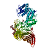
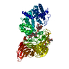
| Entry | Database: PDB / ID: 2v9y | ||||||
|---|---|---|---|---|---|---|---|
| Title | Human aminoimidazole ribonucleotide synthetase | ||||||
 Components Components | PHOSPHORIBOSYLFORMYLGLYCINAMIDINE CYCLO-LIGASE | ||||||
 Keywords Keywords | LIGASE / MULTIFUNCTIONAL ENZYME / STRUCTURAL GENOMICS CONSORTIUM / NUCLEOTIDE-BINDING / PURINE BIOSYNTHESIS / SGC / AIRS / GART / TRANSFERASE / ATP-BINDING / AMINOIMIDAZOLE RIBONUCLEOTIDE SYNTHETASE / PHOSPHORYLATION / PURINE METABOLISM / STRUCTURAL GENOMICS | ||||||
| Function / homology |  Function and homology information Function and homology informationphosphoribosylamine-glycine ligase / phosphoribosylglycinamide formyltransferase 1 / adenine biosynthetic process / phosphoribosylformylglycinamidine cyclo-ligase / phosphoribosylformylglycinamidine cyclo-ligase activity / phosphoribosylglycinamide formyltransferase activity / purine ribonucleoside monophosphate biosynthetic process / phosphoribosylamine-glycine ligase activity / brainstem development / 'de novo' XMP biosynthetic process ...phosphoribosylamine-glycine ligase / phosphoribosylglycinamide formyltransferase 1 / adenine biosynthetic process / phosphoribosylformylglycinamidine cyclo-ligase / phosphoribosylformylglycinamidine cyclo-ligase activity / phosphoribosylglycinamide formyltransferase activity / purine ribonucleoside monophosphate biosynthetic process / phosphoribosylamine-glycine ligase activity / brainstem development / 'de novo' XMP biosynthetic process / Purine ribonucleoside monophosphate biosynthesis / 'de novo' AMP biosynthetic process / purine nucleotide biosynthetic process / GMP biosynthetic process / 'de novo' IMP biosynthetic process / cerebellum development / cerebral cortex development / extracellular exosome / ATP binding / metal ion binding / cytosol Similarity search - Function | ||||||
| Biological species |  HOMO SAPIENS (human) HOMO SAPIENS (human) | ||||||
| Method |  X-RAY DIFFRACTION / SYNCHROTRON / MOLECULAR REPLACEMENT / Resolution: 2.1 Å X-RAY DIFFRACTION / SYNCHROTRON / MOLECULAR REPLACEMENT / Resolution: 2.1 Å | ||||||
 Authors Authors | Welin, M. / Lehtio, L. / Arrowsmith, C.H. / Berglund, H. / Busam, R. / Collins, R. / Dahlgren, L.G. / Herman, M.D. / Edwards, A.M. / Flodin, S. ...Welin, M. / Lehtio, L. / Arrowsmith, C.H. / Berglund, H. / Busam, R. / Collins, R. / Dahlgren, L.G. / Herman, M.D. / Edwards, A.M. / Flodin, S. / Flores, A. / Graslund, S. / Hammarstrom, M. / Hallberg, B.M. / Holmberg-Schiavone, L. / Johansson, I. / Kallas, A. / Karlberg, T. / Kotenyova, T. / Moche, M. / Nyman, T. / Persson, C. / Sagemark, J. / Stenmark, P. / Sundstrom, M. / Thorsell, A.G. / Tresaugues, L. / van den Berg, S. / Weigelt, J. / Nordlund, P. / Structural Genomics Consortium (SGC) | ||||||
 Citation Citation | Journal: Nucleic Acids Res. / Year: 2010 Title: Structural Studies of Tri-Functional Human Gart. Authors: Welin, M. / Grossmann, J.G. / Flodin, S. / Nyman, T. / Stenmark, P. / Tresaugues, L. / Kotenyova, T. / Johansson, I. / Nordlund, P. / Lehtio, L. | ||||||
| History |
|
- Structure visualization
| Structure viewer | Molecule: MolmilJmol/JSmol |
|---|
- Downloads & links
-Download
| PDBx/mmCIF format | 2v9y.cif.gz | 132.5 KB | Display | PDBx/mmCIF format |
|---|---|---|---|---|
| PDB format | pdb2v9y.ent.gz | 103.3 KB | Display | PDB format |
| PDBx/mmJSON format | 2v9y.json.gz | Tree view | PDBx/mmJSON format | |
| Others |  Other downloads Other downloads |
-Validation report
| Arichive directory | https://data.pdbj.org/pub/pdb/validation_reports/v9/2v9yftp://data.pdbj.org/pub/pdb/validation_reports/v9/2v9y | HTTPS FTP |
|---|
-Related structure data
| Related structure data |  2qk4C  1cliS S: Starting model for refinement C: citing same article ( |
|---|---|
| Similar structure data |


-Links
 PDBj
PDBj


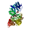
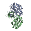
- Assembly
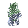
| Deposited unit | 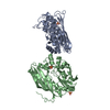
| ||||||||
|---|---|---|---|---|---|---|---|---|---|
| 1 | 
| ||||||||
| 2 | 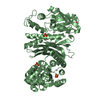
| ||||||||
| Unit cell |
| ||||||||
| Noncrystallographic symmetry (NCS) | NCS oper: (Code: given Matrix: (0.02232, 0.9998, 0.001274), Vector: |
-Components
| #1: Protein | Mass: 35830.016 Da / Num. of mol.: 2 / Fragment: RESIDUES 467-794 / Mutation: YES Source method: isolated from a genetically manipulated source Source: (gene. exp.) HOMO SAPIENS (human) / Plasmid: PNIC-BSA4 / Production host:  References: UniProt: P22102, phosphoribosylformylglycinamidine cyclo-ligase #2: Chemical |   Mass: 96.063 Da / Num. of mol.: 3 / Source method: obtained synthetically / Formula: SO4 Mass: 96.063 Da / Num. of mol.: 3 / Source method: obtained synthetically / Formula: SO4#3: Water | ChemComp-HOH / |  Mass: 18.015 Da / Num. of mol.: 170 / Source method: isolated from a natural source / Formula: H2O Mass: 18.015 Da / Num. of mol.: 170 / Source method: isolated from a natural source / Formula: H2OCompound details | ENGINEERED | Nonpolymer details | SULFATE ION (SO4): FLEXIBLE REGION BETWEEN RESIDUE 770 AND 773 IN BOTH A AND B. | Sequence details | SEQUENCE CONTAINED AN N-TERMINAL HEXAHISTID | |
|---|
-Experimental details
-Experiment
| Experiment | Method: X-RAY DIFFRACTION / Number of used crystals: 1 |
|---|
- Sample preparation
| Crystal | Density Matthews: 2.29 Å3/Da / Density % sol: 46.4 % / Description: NONE |
|---|---|
| Crystal grow | pH: 5.2 Details: 0.1 M BIS-TRIS PH 5.2, 27% PEG 3350, 0.2 M AMMONIUM SULFATE, 0.2 MG/ML OF CHYMOTRYPSIN WAS ADDED PRIOR TO CRYSTALLISATION. |
-Data collection
| Diffraction | Mean temperature: 100 K |
|---|---|
| Diffraction source | Source: SYNCHROTRON / Site: BESSY  / Beamline: 14.1 / Wavelength: 1 / Beamline: 14.1 / Wavelength: 1 |
| Detector | Type: MARRESEARCH / Detector: CCD / Date: Jun 12, 2007 / Details: MIRRORS |
| Radiation | Monochromator: DOUBLE CRYSTAL / Protocol: SINGLE WAVELENGTH / Monochromatic (M) / Laue (L): M / Scattering type: x-ray |
| Radiation wavelength | Wavelength: 1 Å / Relative weight: 1 |
| Reflection | Resolution: 2.1→15 Å / Num. obs: 38171 / % possible obs: 99.6 % / Observed criterion σ(I): 2 / Redundancy: 14.8 % / Rmerge(I) obs: 0.08 / Net I/σ(I): 28.7 |
| Reflection shell | Resolution: 2.1→2.2 Å / Redundancy: 14.9 % / Rmerge(I) obs: 0.39 / Mean I/σ(I) obs: 8.06 / % possible all: 100 |
- Processing
| Software |
| ||||||||||||||||||||||||||||||||||||||||||||||||||||||||||||||||||||||||||||||||||||||||||||||||||||||||||||||||||||||||||||||||||||||||||||||||||||||||||||||||||||||||||||||||||||||
|---|---|---|---|---|---|---|---|---|---|---|---|---|---|---|---|---|---|---|---|---|---|---|---|---|---|---|---|---|---|---|---|---|---|---|---|---|---|---|---|---|---|---|---|---|---|---|---|---|---|---|---|---|---|---|---|---|---|---|---|---|---|---|---|---|---|---|---|---|---|---|---|---|---|---|---|---|---|---|---|---|---|---|---|---|---|---|---|---|---|---|---|---|---|---|---|---|---|---|---|---|---|---|---|---|---|---|---|---|---|---|---|---|---|---|---|---|---|---|---|---|---|---|---|---|---|---|---|---|---|---|---|---|---|---|---|---|---|---|---|---|---|---|---|---|---|---|---|---|---|---|---|---|---|---|---|---|---|---|---|---|---|---|---|---|---|---|---|---|---|---|---|---|---|---|---|---|---|---|---|---|---|---|---|
| Refinement | Method to determine structure: MOLECULAR REPLACEMENT Starting model: PDB ENTRY 1CLI Resolution: 2.1→14.99 Å / Cor.coef. Fo:Fc: 0.942 / Cor.coef. Fo:Fc free: 0.916 / SU B: 9.202 / SU ML: 0.125 / TLS residual ADP flag: LIKELY RESIDUAL / Cross valid method: THROUGHOUT / ESU R: 0.221 / ESU R Free: 0.188 / Stereochemistry target values: MAXIMUM LIKELIHOOD Details: HYDROGENS HAVE BEEN ADDED IN THE RIDING POSITIONS. THE PROTEIN WAS CO CRYSTALLIZED WITH CHYMOTRYPSIN AND THE MOST PROBABLE SEQUENCE IN THE CRYSTAL IS KVDLGGFAGLFDLKAAGFKDPLLA SGTDGVGTKL ...Details: HYDROGENS HAVE BEEN ADDED IN THE RIDING POSITIONS. THE PROTEIN WAS CO CRYSTALLIZED WITH CHYMOTRYPSIN AND THE MOST PROBABLE SEQUENCE IN THE CRYSTAL IS KVDLGGFAGLFDLKAAGFKDPLLA SGTDGVGTKL KIAQLCNKHD TIGQDLVAMC VNDILAQGAE PLFFLDYFSC GKLDLSVTEA VVAGIAKACG KAGCALLGGE TAEMPDMYPP GEYDLAGFAV GAMERDQKLP HLERITEGDV VVGIASSGLH SNGFSLVRKI VAKSSLQYSS PAPDGCGDQT LGDLLLTPTR IYSHSLLPVL RSGHVKAFAH ITGGGLLENI PRVLPEKLGV DLDAQTWRIP RVFSWLQQEG HLSEEEMART FNCGVGAVLV VSKEQTEQIL RGIQQHKEEA WVIGSVVARA EGSPRVKVKN LIESMQINGS VLKN
| ||||||||||||||||||||||||||||||||||||||||||||||||||||||||||||||||||||||||||||||||||||||||||||||||||||||||||||||||||||||||||||||||||||||||||||||||||||||||||||||||||||||||||||||||||||||
| Solvent computation | Ion probe radii: 0.8 Å / Shrinkage radii: 0.8 Å / VDW probe radii: 1.2 Å / Solvent model: MASK | ||||||||||||||||||||||||||||||||||||||||||||||||||||||||||||||||||||||||||||||||||||||||||||||||||||||||||||||||||||||||||||||||||||||||||||||||||||||||||||||||||||||||||||||||||||||
| Displacement parameters | Biso mean: 32.97 Å2
| ||||||||||||||||||||||||||||||||||||||||||||||||||||||||||||||||||||||||||||||||||||||||||||||||||||||||||||||||||||||||||||||||||||||||||||||||||||||||||||||||||||||||||||||||||||||
| Refinement step | Cycle: LAST / Resolution: 2.1→14.99 Å
| ||||||||||||||||||||||||||||||||||||||||||||||||||||||||||||||||||||||||||||||||||||||||||||||||||||||||||||||||||||||||||||||||||||||||||||||||||||||||||||||||||||||||||||||||||||||
| Refine LS restraints |
|