[English] 日本語
Yorodumi
- PDB-1zly: The structure of human glycinamide ribonucleotide transformylase ... -

+
Open data


ID or keywords:

Loading...

-
Basic information

Entry
Database: PDB / ID: 1zly
Title
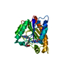
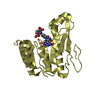
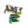
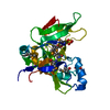
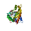
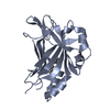
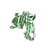
The structure of human glycinamide ribonucleotide transformylase in complex with alpha,beta-N-(hydroxyacetyl)-D-ribofuranosylamine and 10-formyl-5,8,dideazafolate
ComponentsPhosphoribosylglycinamide formyltransferase
KeywordsTRANSFERASE / Purine biosynthesis
Function / homology
Function and homology information


phosphoribosylformylglycinamidine cyclo-ligase / phosphoribosylformylglycinamidine cyclo-ligase activity / adenine biosynthetic process / phosphoribosylamine-glycine ligase / phosphoribosylamine-glycine ligase activity / phosphoribosylglycinamide formyltransferase 1 / purine ribonucleoside monophosphate biosynthetic process / phosphoribosylglycinamide formyltransferase activity / 'de novo' XMP biosynthetic process / brainstem development ...phosphoribosylformylglycinamidine cyclo-ligase / phosphoribosylformylglycinamidine cyclo-ligase activity / adenine biosynthetic process / phosphoribosylamine-glycine ligase / phosphoribosylamine-glycine ligase activity / phosphoribosylglycinamide formyltransferase 1 / purine ribonucleoside monophosphate biosynthetic process / phosphoribosylglycinamide formyltransferase activity / 'de novo' XMP biosynthetic process / brainstem development / Purine ribonucleoside monophosphate biosynthesis / glycine metabolic process / 'de novo' AMP biosynthetic process / GMP biosynthetic process / purine nucleotide biosynthetic process / 'de novo' IMP biosynthetic process / : / tetrahydrofolate biosynthetic process / cerebellum development / : / cerebral cortex development / extracellular exosome / ATP binding / metal ion binding / cytosol
Similarity search - Function
Phosphoribosylformylglycinamidine cyclo-ligase / Phosphoribosylglycinamide synthetase, ATP-grasp (A) domain / Phosphoribosylglycinamide synthetase / Phosphoribosylglycinamide synthetase, conserved site / Phosphoribosylglycinamide synthetase, C-domain / Phosphoribosylglycinamide synthetase, N-terminal / Phosphoribosylglycinamide synthetase, C-domain superfamily / Phosphoribosylglycinamide synthetase, C domain / Phosphoribosylglycinamide synthetase, N domain / Phosphoribosylglycinamide synthetase signature. ...Phosphoribosylformylglycinamidine cyclo-ligase / Phosphoribosylglycinamide synthetase, ATP-grasp (A) domain / Phosphoribosylglycinamide synthetase / Phosphoribosylglycinamide synthetase, conserved site / Phosphoribosylglycinamide synthetase, C-domain / Phosphoribosylglycinamide synthetase, N-terminal / Phosphoribosylglycinamide synthetase, C-domain superfamily / Phosphoribosylglycinamide synthetase, C domain / Phosphoribosylglycinamide synthetase, N domain / Phosphoribosylglycinamide synthetase signature. / Phosphoribosylglycinamide synthetase, C domain / Phosphoribosylglycinamide synthetase, ATP-grasp (A) domain / Phosphoribosylglycinamide synthetase, ATP-grasp (A) domain / Phosphoribosylglycinamide formyltransferase / Formyl transferase, N-terminal domain / PurM-like, N-terminal domain / AIR synthase related protein, N-terminal domain / Phosphoribosylglycinamide formyltransferase, active site / Phosphoribosylglycinamide formyltransferase active site. / PurM-like, C-terminal domain / PurM-like, C-terminal domain superfamily / PurM-like, N-terminal domain superfamily / AIR synthase related protein, C-terminal domain / Formyl transferase, N-terminal / Formyl transferase / Formyl transferase, N-terminal domain superfamily / Rudiment single hybrid motif / ATP-grasp fold, subdomain 1 / Pre-ATP-grasp domain superfamily / ATP-grasp fold / ATP-grasp fold profile. / Rossmann fold / 3-Layer(aba) Sandwich / Alpha Beta
Similarity search - Domain/homology
Chem-DQB / 5-O-phosphono-beta-D-ribofuranosylamine / Trifunctional purine biosynthetic protein adenosine-3
Similarity search - Component
Biological speciesHomo sapiens (human)
MethodX-RAY DIFFRACTION / SYNCHROTRON / MOLECULAR REPLACEMENT / Resolution: 2.07 Å
AuthorsDahms, T.E.S. / Sainz, G. / Giroux, E.L. / Caperelli, C.A. / Smith, J.L.
CitationJournal: Biochemistry / Year: 2005
Title: The apo and ternary complex structures of a chemotherapeutic target: human glycinamide ribonucleotide transformylase.
Authors: Dahms, T.E. / Sainz, G. / Giroux, E.L. / Caperelli, C.A. / Smith, J.L.
History
DepositionMay 9, 2005Deposition site: RCSB / Processing site: RCSB
Revision 1.0Aug 23, 2005Provider: repository / Type: Initial release
Revision 1.1Apr 30, 2008Group: Version format compliance
Revision 1.2Jul 13, 2011Group: Version format compliance
Revision 2.0Jul 29, 2020Group: Atomic model / Derived calculations / Structure summary
Category: atom_site / chem_comp ...atom_site / chem_comp / entity / pdbx_entity_nonpoly / struct_site / struct_site_gen
Item: _atom_site.auth_atom_id / _atom_site.label_atom_id ..._atom_site.auth_atom_id / _atom_site.label_atom_id / _chem_comp.name / _chem_comp.type / _entity.pdbx_description / _pdbx_entity_nonpoly.name
Description: Carbohydrate remediation / Provider: repository / Type: Remediation
Revision 2.1Aug 23, 2023Group: Data collection / Database references ...Data collection / Database references / Refinement description / Structure summary
Category: chem_comp / chem_comp_atom ...chem_comp / chem_comp_atom / chem_comp_bond / database_2 / pdbx_initial_refinement_model
Item: _chem_comp.pdbx_synonyms / _database_2.pdbx_DOI / _database_2.pdbx_database_accession

-
Structure visualization

Structure viewerMolecule:
MolmilJmol/JSmol

Downloads & links

-
Assembly

Deposited unit
A: Phosphoribosylglycinamide formyltransferase
hetero molecules


Theoretical massNumber of molelcules
Total (without water)22,4753
Polymers21,8501
Non-polymers6252
Water4,558253
1


  • Idetical with deposited unit
  • defined by author
TypeNameSymmetry operationNumber
identity operation1_555x,y,z1
Unit cell
Length a, b, c (Å)75.900, 75.900, 101.000
Angle α, β, γ (deg.)90.00, 90.00, 120.00
Int Tables number154
Space group name H-MP3221

-
Components

#1: Protein Phosphoribosylglycinamide formyltransferase / GART / GAR transformylase / 5'-phosphoribosylglycinamide transformylase


Mass: 21850.061 Da / Num. of mol.: 1 / Fragment: residues 808-1010, GART
Source method: isolated from a genetically manipulated source
Source: (gene. exp.) Homo sapiens (human) / Gene: GART, PRGS / Plasmid: pET17b / Production host: Escherichia coli (E. coli) / Strain (production host): BL21(DE3) pLysS
References: UniProt: P22102, phosphoribosylglycinamide formyltransferase 1
#2: Sugar ChemComp-GRF / 5-O-phosphono-beta-D-ribofuranosylamine / 5-O-phosphono-beta-D-ribosylamine / 5-O-phosphono-D-ribosylamine / 5-O-phosphono-ribosylamine


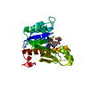
Type: D-saccharide, beta linking / Mass: 229.125 Da / Num. of mol.: 1 / Source method: obtained synthetically / Formula: C5H12NO7P
#3: Chemical ChemComp-DQB / 4-[(4-{[(2-AMINO-4-OXO-3,4-DIHYDROQUINAZOLIN-6-YL)METHYL]AMINO}BENZOYL)AMINO]BUTANOIC ACID


Mass: 395.412 Da / Num. of mol.: 1 / Source method: obtained synthetically / Formula: C20H21N5O4
#4: Water ChemComp-HOH / water


Mass: 18.015 Da / Num. of mol.: 253 / Source method: isolated from a natural source / Formula: H2O

-
Experimental details

-
Experiment

ExperimentMethod: X-RAY DIFFRACTION / Number of used crystals: 1

-
Sample preparation

Crystal
IDDensity Matthews3/Da)Density % sol (%)
13.773
2
Crystal grow
Temperature (K)Crystal-IDMethodpHDetails
3001vapor diffusion, hanging drop65 mg/ml protein in 10 mM Hepes pH 7.5, 1 mM DTE 1:1 with well solution (100 mM Na citrate pH 6.0, 20% 2-propanol (v/v), 20% PEG-4000 (w/v)) , VAPOR DIFFUSION, HANGING DROP, temperature 300K
3002vapor diffusion, sitting drop6.35 mg/ml protein in 10 mM Hepes pH 7.5, 1 mM DTE 1:1 with well solution (100 mM Na citrate pH 6.0, 6-8% 2-propanol (v/v), 16-18% PEG-4000 (w/v)) , pH 6.3, VAPOR DIFFUSION, SITTING DROP, temperature 300K

-
Data collection

Diffraction
IDMean temperature (K)Crystal-ID
12821
21101
Diffraction source
SourceSiteBeamlineTypeIDWavelength (Å)
ROTATING ANODERIGAKU RU20011.5418
SYNCHROTRONCHESS F220.68
Detector
TypeIDDetectorDate
RIGAKU RAXIS IIC1IMAGE PLATEFeb 25, 1997
ADSC QUANTUM 42CCDAug 12, 1997
Radiation
IDMonochromatorProtocolMonochromatic (M) / Laue (L)Scattering typeWavelength-ID
1GRAPHITESINGLE WAVELENGTHMx-ray1
2Si 111 CHANNELSINGLE WAVELENGTHMx-ray1
Radiation wavelength
IDWavelength (Å)Relative weight
11.54181
20.681
ReflectionResolution: 2.07→40 Å / Num. all: 21205 / Num. obs: 19915 / % possible obs: 99.5 % / Observed criterion σ(F): 2 / Observed criterion σ(I): 1 / Redundancy: 3.9 % / Biso Wilson estimate: 53.8 Å2 / Rsym value: 0.073 / Net I/σ(I): 7.7
Reflection shellResolution: 2.07→2.17 Å / Mean I/σ(I) obs: 3.7 / Rsym value: 0.296 / % possible all: 3

-
Processing

Software
NameVersionClassification
DENZOdata reduction
SCALEPACKdata scaling
AMoREphasing
X-PLOR3.1refinement
RefinementMethod to determine structure: MOLECULAR REPLACEMENT
Starting model: 1ZLX
Resolution: 2.07→40 Å / Isotropic thermal model: Anisotropic / σ(F): 0 / σ(I): 2 / Stereochemistry target values: Engh & Huber
RfactorNum. reflection% reflectionSelection details
Rfree0.236 1015 -Selected
Rwork0.19 ---
all0.222 19915 --
obs0.2 18900 95 %-
Displacement parametersBiso mean: 53.8 Å2
Baniso -1Baniso -2Baniso -3
1--5.79 Å2-3.49 Å2-
2---5.79 Å2-
3---11.59 Å2
Refine analyzeLuzzati coordinate error free: 0.28 Å
Refinement stepCycle: LAST / Resolution: 2.07→40 Å
ProteinNucleic acidLigandSolventTotal
Num. atoms1507 0 43 253 1803
Refine LS restraints
Refine-IDTypeDev ideal
X-RAY DIFFRACTIONc_bond_d0.006
X-RAY DIFFRACTIONc_angle_deg1.45
X-RAY DIFFRACTIONc_mcbond_it2.94

+
About Yorodumi

-
News

-
Feb 9, 2022. New format data for meta-information of EMDB entries

New format data for meta-information of EMDB entries

  • Version 3 of the EMDB header file is now the official format.
  • The previous official version 1.9 will be removed from the archive.

Related info.:EMDB header

External links:wwPDB to switch to version 3 of the EMDB data model

-
Aug 12, 2020. Covid-19 info

Covid-19 info

URL: https://pdbjlvh1.pdbj.org/emnavi/covid19.php

New page: Covid-19 featured information page in EM Navigator.

Related info.:Covid-19 info / Mar 5, 2020. Novel coronavirus structure data

+
Mar 5, 2020. Novel coronavirus structure data

Novel coronavirus structure data

Related info.:Yorodumi Speices / Aug 12, 2020. Covid-19 info

External links:COVID-19 featured content - PDBj / Molecule of the Month (242):Coronavirus Proteases

+
Jan 31, 2019. EMDB accession codes are about to change! (news from PDBe EMDB page)

EMDB accession codes are about to change! (news from PDBe EMDB page)

  • The allocation of 4 digits for EMDB accession codes will soon come to an end. Whilst these codes will remain in use, new EMDB accession codes will include an additional digit and will expand incrementally as the available range of codes is exhausted. The current 4-digit format prefixed with “EMD-” (i.e. EMD-XXXX) will advance to a 5-digit format (i.e. EMD-XXXXX), and so on. It is currently estimated that the 4-digit codes will be depleted around Spring 2019, at which point the 5-digit format will come into force.
  • The EM Navigator/Yorodumi systems omit the EMD- prefix.

Related info.:Q: What is EMD? / ID/Accession-code notation in Yorodumi/EM Navigator

External links:EMDB Accession Codes are Changing Soon! / Contact to PDBj

+
Jul 12, 2017. Major update of PDB

Major update of PDB

  • wwPDB released updated PDB data conforming to the new PDBx/mmCIF dictionary.
  • This is a major update changing the version number from 4 to 5, and with Remediation, in which all the entries are updated.
  • In this update, many items about electron microscopy experimental information are reorganized (e.g. em_software).
  • Now, EM Navigator and Yorodumi are based on the updated data.

External links:wwPDB Remediation / Enriched Model Files Conforming to OneDep Data Standards Now Available in the PDB FTP Archive

-
Yorodumi

Thousand views of thousand structures

  • Yorodumi is a browser for structure data from EMDB, PDB, SASBDB, etc.
  • This page is also the successor to EM Navigator detail page, and also detail information page/front-end page for Omokage search.
  • The word "yorodu" (or yorozu) is an old Japanese word meaning "ten thousand". "mi" (miru) is to see.

Related info.:EMDB / PDB / SASBDB / Comparison of 3 databanks / Yorodumi Search / Aug 31, 2016. New EM Navigator & Yorodumi / Yorodumi Papers / Jmol/JSmol / Function and homology information / Changes in new EM Navigator and Yorodumi

Read more