 Movie
Movie Controller

[English] 日本語
 Yorodumi
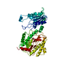
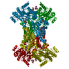
- PDB-2v3z: Glu383Ala Escherichia coli aminopeptidase P in complex with substrate -
+ Open data

- Basic information
| Entry | Database: PDB / ID: 2v3z | ||||||
|---|---|---|---|---|---|---|---|
| Title | Glu383Ala Escherichia coli aminopeptidase P in complex with substrate | ||||||
 Components Components |
| ||||||
 Keywords Keywords | HYDROLASE / 'PITA-BREAD' ENZYME / PROLINE- SPECIFIC ENZYME / AMINOPEPTIDASE P / MANGANESE ENZYME / PROTEASE / MANGANESE / METAL-BINDING / METALLOENZYME / AMINOPEPTIDASE / METALLOPROTEASE | ||||||
| Function / homology |  Function and homology information Function and homology informationXaa-Pro aminopeptidase / metalloexopeptidase activity / metalloaminopeptidase activity / aminopeptidase activity / manganese ion binding / protein homotetramerization / protein-containing complex / proteolysis / identical protein binding / cytosol Similarity search - Function | ||||||
| Biological species |  | ||||||
| Method |  X-RAY DIFFRACTION / SYNCHROTRON / MOLECULAR REPLACEMENT / Resolution: 1.56 Å X-RAY DIFFRACTION / SYNCHROTRON / MOLECULAR REPLACEMENT / Resolution: 1.56 Å | ||||||
 Authors Authors | Graham, S.C. / Guss, J.M. | ||||||
 Citation Citation | Journal: Arch.Biochem.Biophys. / Year: 2008 Title: Complexes of Mutants of Escherichia Coli Aminopeptidase P and the Tripeptide Substrate Valproleu. Authors: Graham, S.C. / Guss, J.M. | ||||||
| History |
|
- Structure visualization
| Structure viewer | Molecule: MolmilJmol/JSmol |
|---|
- Downloads & links
-Download
| PDBx/mmCIF format | 2v3z.cif.gz | 121.8 KB | Display | PDBx/mmCIF format |
|---|---|---|---|---|
| PDB format | pdb2v3z.ent.gz | 94.6 KB | Display | PDB format |
| PDBx/mmJSON format | 2v3z.json.gz | Tree view | PDBx/mmJSON format | |
| Others |  Other downloads Other downloads |
-Validation report
| Arichive directory | https://data.pdbj.org/pub/pdb/validation_reports/v3/2v3zftp://data.pdbj.org/pub/pdb/validation_reports/v3/2v3z | HTTPS FTP |
|---|
-Related structure data
| Related structure data |  2v3xC  2v3yC  1wl9S S: Starting model for refinement C: citing same article ( |
|---|---|
| Similar structure data |


-Links
 PDBj
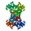
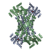
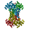
PDBj- Assembly
| Deposited unit | 
| ||||||||
|---|---|---|---|---|---|---|---|---|---|
| 1 | 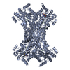
| ||||||||
| Unit cell |
|
-Components
| #1: Protein | Mass: 49702.027 Da / Num. of mol.: 1 / Mutation: YES Source method: isolated from a genetically manipulated source Source: (gene. exp.) | ||||||||||
|---|---|---|---|---|---|---|---|---|---|---|---|
| #2: Protein/peptide | Mass: 327.419 Da / Num. of mol.: 1 / Source method: obtained synthetically / Details: TRIPEPTIDE SUBSTRATE OF AMINOPEPTIDASE P / Source: (synth.) | ||||||||||
| #3: Chemical |   Mass: 54.938 Da / Num. of mol.: 2 / Source method: obtained synthetically / Formula: Mn Mass: 54.938 Da / Num. of mol.: 2 / Source method: obtained synthetically / Formula: Mn#4: Chemical | ChemComp-CL / |   Mass: 35.453 Da / Num. of mol.: 1 / Source method: obtained synthetically / Formula: Cl Mass: 35.453 Da / Num. of mol.: 1 / Source method: obtained synthetically / Formula: Cl#5: Water | ChemComp-HOH / |  Mass: 18.015 Da / Num. of mol.: 731 / Source method: isolated from a natural source / Formula: H2O Mass: 18.015 Da / Num. of mol.: 731 / Source method: isolated from a natural source / Formula: H2OCompound details | ENGINEERED RESIDUE IN CHAIN A, GLU 383 TO ALA RELEASE OF ANY N-TERMINAL AMINO ACID, INCLUDING ...ENGINEERED | Has protein modification | Y | Sequence details | E383A MUTANT | |
-Experimental details
-Experiment
| Experiment | Method: X-RAY DIFFRACTION / Number of used crystals: 1 |
|---|
- Sample preparation
| Crystal | Density Matthews: 4.4 Å3/Da / Density % sol: 72.1 % Description: STARTING MODEL WAS STRIPPED OF LIGANDS, SOLVENT AND ALTERNATE CONFORMERS BEFORE REFINEMENT |
|---|---|
| Crystal grow | Temperature: 277 K / Method: vapor diffusion, hanging drop / pH: 8.5 Details: CRYSTALLISED IN 30% PEG 4K, 0.1 M TRIS PH 8.8 AT 277K. SOAKED IN 30% PEG 4K, 0.1 M TRIS PH 8.5, 1 MM MNCL2, 5 MM VAL-PRO-LEU, 10% MPD FOR 30 MIN AT 277K IMMEDIATELY PRIOR TO DATA COLLECTION. |
-Data collection
| Diffraction | Mean temperature: 100 K |
|---|---|
| Diffraction source | Source: SYNCHROTRON / Site: APS  / Beamline: 23-ID-B / Wavelength: 0.97934 / Beamline: 23-ID-B / Wavelength: 0.97934 |
| Detector | Type: ADSC CCD / Detector: CCD / Date: Apr 22, 2006 / Details: MIRRORS |
| Radiation | Monochromator: SILICON 111 / Protocol: SINGLE WAVELENGTH / Monochromatic (M) / Laue (L): M / Scattering type: x-ray |
| Radiation wavelength | Wavelength: 0.97934 Å / Relative weight: 1 |
| Reflection | Resolution: 1.56→50 Å / Num. obs: 125677 / % possible obs: 99.4 % / Observed criterion σ(I): -3 / Redundancy: 11 % / Biso Wilson estimate: 17.78 Å2 / Rmerge(I) obs: 0.07 / Net I/σ(I): 32.5 |
| Reflection shell | Resolution: 1.56→1.62 Å / Redundancy: 10 % / Rmerge(I) obs: 0.58 / Mean I/σ(I) obs: 4.3 / % possible all: 99.8 |
- Processing
| Software |
| ||||||||||||||||||||||||||||||||||||||||||||||||||||||||||||||||||||||||||||||||||||||||||||||||||||||||||||||||||||||||||||||||||||||||||||||||||||||||||||||||||||||||||||||||||||||
|---|---|---|---|---|---|---|---|---|---|---|---|---|---|---|---|---|---|---|---|---|---|---|---|---|---|---|---|---|---|---|---|---|---|---|---|---|---|---|---|---|---|---|---|---|---|---|---|---|---|---|---|---|---|---|---|---|---|---|---|---|---|---|---|---|---|---|---|---|---|---|---|---|---|---|---|---|---|---|---|---|---|---|---|---|---|---|---|---|---|---|---|---|---|---|---|---|---|---|---|---|---|---|---|---|---|---|---|---|---|---|---|---|---|---|---|---|---|---|---|---|---|---|---|---|---|---|---|---|---|---|---|---|---|---|---|---|---|---|---|---|---|---|---|---|---|---|---|---|---|---|---|---|---|---|---|---|---|---|---|---|---|---|---|---|---|---|---|---|---|---|---|---|---|---|---|---|---|---|---|---|---|---|---|
| Refinement | Method to determine structure: MOLECULAR REPLACEMENT Starting model: PDB ENTRY 1WL9 Resolution: 1.56→29.66 Å / Cor.coef. Fo:Fc: 0.975 / Cor.coef. Fo:Fc free: 0.97 / SU B: 1.35 / SU ML: 0.026 / TLS residual ADP flag: LIKELY RESIDUAL / Cross valid method: THROUGHOUT / ESU R: 0.045 / ESU R Free: 0.045 / Stereochemistry target values: MAXIMUM LIKELIHOOD / Details: HYDROGENS HAVE BEEN ADDED IN THE RIDING POSITIONS.
| ||||||||||||||||||||||||||||||||||||||||||||||||||||||||||||||||||||||||||||||||||||||||||||||||||||||||||||||||||||||||||||||||||||||||||||||||||||||||||||||||||||||||||||||||||||||
| Solvent computation | Ion probe radii: 0.8 Å / Shrinkage radii: 0.8 Å / VDW probe radii: 1.4 Å / Solvent model: MASK | ||||||||||||||||||||||||||||||||||||||||||||||||||||||||||||||||||||||||||||||||||||||||||||||||||||||||||||||||||||||||||||||||||||||||||||||||||||||||||||||||||||||||||||||||||||||
| Displacement parameters | Biso mean: 10.84 Å2
| ||||||||||||||||||||||||||||||||||||||||||||||||||||||||||||||||||||||||||||||||||||||||||||||||||||||||||||||||||||||||||||||||||||||||||||||||||||||||||||||||||||||||||||||||||||||
| Refinement step | Cycle: LAST / Resolution: 1.56→29.66 Å
| ||||||||||||||||||||||||||||||||||||||||||||||||||||||||||||||||||||||||||||||||||||||||||||||||||||||||||||||||||||||||||||||||||||||||||||||||||||||||||||||||||||||||||||||||||||||
| Refine LS restraints |
|