 Movie
Movie Controller


+ Open data

- Basic information
| Entry | Database: PDB / ID: 2bwv | ||||||
|---|---|---|---|---|---|---|---|
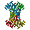
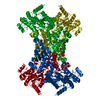
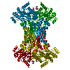
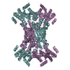
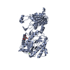
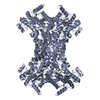
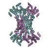
| Title | His361Ala Escherichia coli Aminopeptidase P | ||||||
 Components Components | AMINOPEPTIDASE P | ||||||
 Keywords Keywords | HYDROLASE / METALLOENZYME / 'PITA-BREAD' ENZYME / PROLINE-SPECIFIC ENZYME / MANGANESE ENZYME | ||||||
| Function / homology |  Function and homology information Function and homology informationXaa-Pro aminopeptidase / metalloexopeptidase activity / metalloaminopeptidase activity / aminopeptidase activity / manganese ion binding / protein homotetramerization / protein-containing complex / proteolysis / identical protein binding / cytosol Similarity search - Function | ||||||
| Biological species |  | ||||||
| Method |  X-RAY DIFFRACTION / MOLECULAR REPLACEMENT / Resolution: 1.7 Å X-RAY DIFFRACTION / MOLECULAR REPLACEMENT / Resolution: 1.7 Å | ||||||
 Authors Authors | Graham, S.C. / Guss, J.M. | ||||||
 Citation Citation | Journal: Biochemistry / Year: 2006 Title: Kinetic and Crystallographic Analysis of Mutant Escherichia Coli Aminopeptidase P: Insights Into Substrate Recognition and the Mechanism of Catalysis. Authors: Graham, S.C. / Lilley, P.E. / Lee, M. / Schaeffer, P.M. / Kralicek, A.V. / Dixon, N.E. / Guss, J.M. | ||||||
| History |
|
- Structure visualization
| Structure viewer | Molecule: MolmilJmol/JSmol |
|---|
- Downloads & links
-Download
| PDBx/mmCIF format | 2bwv.cif.gz | 116.6 KB | Display | PDBx/mmCIF format |
|---|---|---|---|---|
| PDB format | pdb2bwv.ent.gz | 89.6 KB | Display | PDB format |
| PDBx/mmJSON format | 2bwv.json.gz | Tree view | PDBx/mmJSON format | |
| Others |  Other downloads Other downloads |
-Validation report
| Arichive directory | https://data.pdbj.org/pub/pdb/validation_reports/bw/2bwvftp://data.pdbj.org/pub/pdb/validation_reports/bw/2bwv | HTTPS FTP |
|---|
-Related structure data
| Related structure data |  2bwsC  2bwtC  2bwuC  2bwwC  2bwxC  2bwyC  1wl9S S: Starting model for refinement C: citing same article ( |
|---|---|
| Similar structure data |


-Links
 PDBj
PDBj- Assembly
| Deposited unit | 
| ||||||||
|---|---|---|---|---|---|---|---|---|---|
| 1 | 
| ||||||||
| Unit cell |
|
-Components
| #1: Protein | Mass: 49692.996 Da / Num. of mol.: 1 / Mutation: YES Source method: isolated from a genetically manipulated source Source: (gene. exp.) | ||||||||
|---|---|---|---|---|---|---|---|---|---|
| #2: Chemical |   Mass: 54.938 Da / Num. of mol.: 2 / Source method: obtained synthetically / Formula: Mn Mass: 54.938 Da / Num. of mol.: 2 / Source method: obtained synthetically / Formula: Mn#3: Chemical | ChemComp-CL / |   Mass: 35.453 Da / Num. of mol.: 1 / Source method: obtained synthetically / Formula: Cl Mass: 35.453 Da / Num. of mol.: 1 / Source method: obtained synthetically / Formula: Cl#4: Water | ChemComp-HOH / |  Mass: 18.015 Da / Num. of mol.: 649 / Source method: isolated from a natural source / Formula: H2O Mass: 18.015 Da / Num. of mol.: 649 / Source method: isolated from a natural source / Formula: H2OCompound details | ENGINEERED | Has protein modification | Y | |
-Experimental details
-Experiment
| Experiment | Method: X-RAY DIFFRACTION / Number of used crystals: 1 |
|---|
- Sample preparation
| Crystal | Density Matthews: 4.4 Å3/Da / Density % sol: 71.9 % Description: STARTING MODEL WAS STRIPPED OF HETATMS AND MULTIPLE CONFORMERS |
|---|---|
| Crystal grow | Temperature: 277 K / Method: vapor diffusion, hanging drop / pH: 8.5 Details: HANGING DROP VAPOUR DIFFUSION AT 4C. 2 UL 6 MG/ML APPRO PLUS 2 UL RESERVOIR SOLUTION: 28 % PEG 4K, 0.1 M TRIS PH 8.5. SOAKED IN RESERVOIR SOLUTION SUPPLEMENTED WITH 10% MPD AND 1 MM MNCL2 ...Details: HANGING DROP VAPOUR DIFFUSION AT 4C. 2 UL 6 MG/ML APPRO PLUS 2 UL RESERVOIR SOLUTION: 28 % PEG 4K, 0.1 M TRIS PH 8.5. SOAKED IN RESERVOIR SOLUTION SUPPLEMENTED WITH 10% MPD AND 1 MM MNCL2 FOR 1 HR AT 4C PRIOR TO CRYOCOOLING. |
-Data collection
| Diffraction | Mean temperature: 100 K |
|---|---|
| Diffraction source | Source: ROTATING ANODE / Type: RIGAKU RU200H / Wavelength: 1.5418 |
| Detector | Type: MARRESEARCH / Detector: IMAGE PLATE / Date: Apr 5, 2005 / Details: OSMIC MIRRORS |
| Radiation | Protocol: SINGLE WAVELENGTH / Monochromatic (M) / Laue (L): M / Scattering type: x-ray |
| Radiation wavelength | Wavelength: 1.5418 Å / Relative weight: 1 |
| Reflection | Resolution: 1.7→65.23 Å / Num. obs: 96986 / % possible obs: 99.4 % / Observed criterion σ(I): 6 / Redundancy: 6 % / Biso Wilson estimate: 22.44 Å2 / Rmerge(I) obs: 0.06 / Net I/σ(I): 19.7 |
| Reflection shell | Resolution: 1.7→1.79 Å / Redundancy: 5.1 % / Rmerge(I) obs: 0.48 / Mean I/σ(I) obs: 2.8 / % possible all: 99.5 |
- Processing
| Software |
| ||||||||||||||||||||||||||||||||||||||||||||||||||||||||||||||||||||||||||||||||||||||||||||||||||||||||||||||||||||||||||||||||||||||||||||||||||||||||||||||||||||||||||||||||||||||
|---|---|---|---|---|---|---|---|---|---|---|---|---|---|---|---|---|---|---|---|---|---|---|---|---|---|---|---|---|---|---|---|---|---|---|---|---|---|---|---|---|---|---|---|---|---|---|---|---|---|---|---|---|---|---|---|---|---|---|---|---|---|---|---|---|---|---|---|---|---|---|---|---|---|---|---|---|---|---|---|---|---|---|---|---|---|---|---|---|---|---|---|---|---|---|---|---|---|---|---|---|---|---|---|---|---|---|---|---|---|---|---|---|---|---|---|---|---|---|---|---|---|---|---|---|---|---|---|---|---|---|---|---|---|---|---|---|---|---|---|---|---|---|---|---|---|---|---|---|---|---|---|---|---|---|---|---|---|---|---|---|---|---|---|---|---|---|---|---|---|---|---|---|---|---|---|---|---|---|---|---|---|---|---|
| Refinement | Method to determine structure: MOLECULAR REPLACEMENT Starting model: PDB ENTRY 1WL9 Resolution: 1.7→154.3 Å / Cor.coef. Fo:Fc: 0.972 / Cor.coef. Fo:Fc free: 0.964 / SU B: 2.188 / SU ML: 0.038 / Cross valid method: THROUGHOUT / ESU R: 0.06 / ESU R Free: 0.06 / Stereochemistry target values: MAXIMUM LIKELIHOOD / Details: HYDROGENS HAVE BEEN ADDED IN THE RIDING POSITIONS.
| ||||||||||||||||||||||||||||||||||||||||||||||||||||||||||||||||||||||||||||||||||||||||||||||||||||||||||||||||||||||||||||||||||||||||||||||||||||||||||||||||||||||||||||||||||||||
| Solvent computation | Ion probe radii: 0.8 Å / Shrinkage radii: 0.8 Å / VDW probe radii: 1.2 Å / Solvent model: MASK | ||||||||||||||||||||||||||||||||||||||||||||||||||||||||||||||||||||||||||||||||||||||||||||||||||||||||||||||||||||||||||||||||||||||||||||||||||||||||||||||||||||||||||||||||||||||
| Displacement parameters | Biso mean: 21.04 Å2
| ||||||||||||||||||||||||||||||||||||||||||||||||||||||||||||||||||||||||||||||||||||||||||||||||||||||||||||||||||||||||||||||||||||||||||||||||||||||||||||||||||||||||||||||||||||||
| Refinement step | Cycle: LAST / Resolution: 1.7→154.3 Å
| ||||||||||||||||||||||||||||||||||||||||||||||||||||||||||||||||||||||||||||||||||||||||||||||||||||||||||||||||||||||||||||||||||||||||||||||||||||||||||||||||||||||||||||||||||||||
| Refine LS restraints |
|