 Movie
Movie Controller


+ Open data

- Basic information
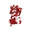
| Entry | Database: PDB / ID: 1n51 | |||||||||
|---|---|---|---|---|---|---|---|---|---|---|
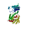
| Title | Aminopeptidase P in complex with the inhibitor apstatin | |||||||||
 Components Components |
| |||||||||
 Keywords Keywords | HYDROLASE/HYDROLASE INHIBITOR / AMINOPEPTIDASE / PROLINE SPECIFIC / MANGANESE ENZYME / HYDROLASE-HYDROLASE INHIBITOR COMPLEX | |||||||||
| Function / homology |  Function and homology information Function and homology informationXaa-Pro aminopeptidase / metalloexopeptidase activity / metalloaminopeptidase activity / aminopeptidase activity / manganese ion binding / protein homotetramerization / protein-containing complex / proteolysis / identical protein binding / cytosol Similarity search - Function | |||||||||
| Biological species |  synthetic (others) | |||||||||
| Method |  X-RAY DIFFRACTION / MOLECULAR REPLACEMENT / Resolution: 2.3 Å X-RAY DIFFRACTION / MOLECULAR REPLACEMENT / Resolution: 2.3 Å | |||||||||
 Authors Authors | Graham, S.C. / Maher, M.J. / Lee, M.H. / Simmons, W.H. / Freeman, H.C. / Guss, J.M. | |||||||||
 Citation Citation | Journal: Acta Crystallogr.,Sect.D / Year: 2004 Title: Structure of Escherichia coli aminopeptidase P in complex with the inhibitor apstatin. Authors: Graham, S.C. / Maher, M.J. / Simmons, W.H. / Freeman, H.C. / Guss, J.M. #1: Journal: Acta Crystallogr.,Sect.D / Year: 1998Title: Crystallography & NMR System: A New Software Suite for Macromolecular Structure Determination Authors: Brunger, A.T. / Adams, P.D. / Clore, G.M. / Delano, W.L. / Gros, P. / Grosse-Kunstleve, R. / Jiang, J.-S. / Kuszewski, J. / Nilges, M. / Pannu, N.S. / Read, R.J. / Rice, L.M. / Simonson, T. / Warren, G. | |||||||||
| History |
|
- Structure visualization
| Structure viewer | Molecule: MolmilJmol/JSmol |
|---|
- Downloads & links
-Download
| PDBx/mmCIF format | 1n51.cif.gz | 113 KB | Display | PDBx/mmCIF format |
|---|---|---|---|---|
| PDB format | pdb1n51.ent.gz | 85.6 KB | Display | PDB format |
| PDBx/mmJSON format | 1n51.json.gz | Tree view | PDBx/mmJSON format | |
| Others |  Other downloads Other downloads |
-Validation report
| Arichive directory | https://data.pdbj.org/pub/pdb/validation_reports/n5/1n51ftp://data.pdbj.org/pub/pdb/validation_reports/n5/1n51 | HTTPS FTP |
|---|
-Related structure data
| Related structure data |  1az9 S: Starting model for refinement |
|---|---|
| Similar structure data |


-Links
 PDBj
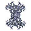
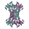
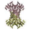
PDBj- Assembly
| Deposited unit | 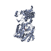
| |||||||||
|---|---|---|---|---|---|---|---|---|---|---|
| 1 | 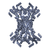
| |||||||||
| 2 | 
| |||||||||
| 3 | 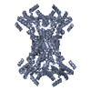
| |||||||||
| Unit cell |
| |||||||||
| Components on special symmetry positions |
| |||||||||
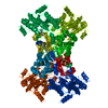
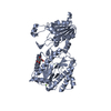
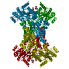
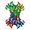
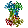
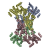
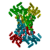
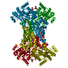
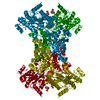
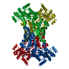
| Details | Biological assembly is a tetramer formed by the crystallographic operations: (X,Y,Z), (-Y+1/2,-X+1/2,-Z+1/2), (Y-1/2,X+1/2,-Z+1/2), (-X,1-Y,Z) |
-Components
| #1: Protein | Mass: 49744.062 Da / Num. of mol.: 1 Source method: isolated from a genetically manipulated source Source: (gene. exp.) | ||
|---|---|---|---|
| #2: Protein/peptide |   Type: Peptide-like / Class: Enzyme inhibitor / Mass: 458.530 Da / Num. of mol.: 1 / Source method: obtained synthetically / Source: (synth.) synthetic (others) / References: APSTATIN Type: Peptide-like / Class: Enzyme inhibitor / Mass: 458.530 Da / Num. of mol.: 1 / Source method: obtained synthetically / Source: (synth.) synthetic (others) / References: APSTATIN | ||
| #3: Chemical |   Mass: 54.938 Da / Num. of mol.: 3 / Source method: obtained synthetically / Formula: Mn Mass: 54.938 Da / Num. of mol.: 3 / Source method: obtained synthetically / Formula: Mn#4: Water | ChemComp-HOH / |  Mass: 18.015 Da / Num. of mol.: 425 / Source method: isolated from a natural source / Formula: H2O Mass: 18.015 Da / Num. of mol.: 425 / Source method: isolated from a natural source / Formula: H2O |
-Experimental details
-Experiment
| Experiment | Method: X-RAY DIFFRACTION / Number of used crystals: 1 |
|---|
- Sample preparation
| Crystal | Density Matthews: 5.02 Å3/Da / Density % sol: 75.28 % | ||||||||||||||||||||||||||||||
|---|---|---|---|---|---|---|---|---|---|---|---|---|---|---|---|---|---|---|---|---|---|---|---|---|---|---|---|---|---|---|---|
| Crystal grow | Temperature: 277 K / Method: vapor diffusion, hanging drop / pH: 6.8 Details: Magnesium Acetate, Sodium Cacodylate, MPD, pH 6.8, VAPOR DIFFUSION, HANGING DROP, temperature 277K | ||||||||||||||||||||||||||||||
| Crystal grow | *PLUS Temperature: 277 K / Method: vapor diffusion, hanging drop | ||||||||||||||||||||||||||||||
| Components of the solutions | *PLUS
|
-Data collection
| Diffraction | Mean temperature: 100 K |
|---|---|
| Diffraction source | Source: ROTATING ANODE / Type: RIGAKU RU200 / Wavelength: 1.5418 |
| Detector | Type: RIGAKU RAXIS IIC / Detector: IMAGE PLATE / Date: Oct 24, 2000 / Details: YALE MIRRORS |
| Radiation | Monochromator: YALE MIRRORS / Protocol: SINGLE WAVELENGTH / Monochromatic (M) / Laue (L): M / Scattering type: x-ray |
| Radiation wavelength | Wavelength: 1.5418 Å / Relative weight: 1 |
| Reflection | Resolution: 2.2→99 Å / Num. all: 48993 / Num. obs: 55327 / % possible obs: 95.5 % / Observed criterion σ(F): 0 / Observed criterion σ(I): 0 / Redundancy: 2.5 % / Biso Wilson estimate: 42.6 Å2 / Limit h max: 63 / Limit h min: 0 / Limit k max: 44 / Limit k min: 0 / Limit l max: 104 / Limit l min: 0 / Observed criterion F max: 3136609.46 / Observed criterion F min: 17.3 / Rmerge(I) obs: 0.046 / Net I/σ(I): 16.58 |
| Reflection shell | Resolution: 2.2→2.28 Å / Redundancy: 1.83 % / Rmerge(I) obs: 0.433 / Mean I/σ(I) obs: 1.46 / Num. unique all: 4729 / % possible all: 83.1 |
| Reflection | *PLUS Highest resolution: 2.3 Å / Lowest resolution: 20 Å / % possible obs: 97 % / Redundancy: 2.6 % / Rmerge(I) obs: 0.044 |
| Reflection shell | *PLUS % possible obs: 96.2 % / Redundancy: 2.4 % / Rmerge(I) obs: 0.411 / Mean I/σ(I) obs: 2 |
- Processing
| Software |
| ||||||||||||||||||||||||||||||||||||||||||||||||||||||||||||||||||||||||||||||||||||||||||
|---|---|---|---|---|---|---|---|---|---|---|---|---|---|---|---|---|---|---|---|---|---|---|---|---|---|---|---|---|---|---|---|---|---|---|---|---|---|---|---|---|---|---|---|---|---|---|---|---|---|---|---|---|---|---|---|---|---|---|---|---|---|---|---|---|---|---|---|---|---|---|---|---|---|---|---|---|---|---|---|---|---|---|---|---|---|---|---|---|---|---|---|
| Refinement | Method to determine structure: MOLECULAR REPLACEMENT Starting model: PDB 1AZ9 1az9 Resolution: 2.3→19.9 Å / Rfactor Rfree error: 0.004 / Occupancy max: 1 / Occupancy min: 0 / Isotropic thermal model: anisotropic / Cross valid method: THROUGHOUT / σ(F): 0 / σ(I): 0 / Stereochemistry target values: Engh & Huber
| ||||||||||||||||||||||||||||||||||||||||||||||||||||||||||||||||||||||||||||||||||||||||||
| Solvent computation | Solvent model: CNS bulk solvent model used / Bsol: 37.434 Å2 / ksol: 0.299544 e/Å3 | ||||||||||||||||||||||||||||||||||||||||||||||||||||||||||||||||||||||||||||||||||||||||||
| Displacement parameters | Biso max: 100.3 Å2 / Biso mean: 44.58 Å2 / Biso min: 18.16 Å2
| ||||||||||||||||||||||||||||||||||||||||||||||||||||||||||||||||||||||||||||||||||||||||||
| Refine analyze |
| ||||||||||||||||||||||||||||||||||||||||||||||||||||||||||||||||||||||||||||||||||||||||||
| Refinement step | Cycle: LAST / Resolution: 2.3→19.9 Å
| ||||||||||||||||||||||||||||||||||||||||||||||||||||||||||||||||||||||||||||||||||||||||||
| Refine LS restraints |
| ||||||||||||||||||||||||||||||||||||||||||||||||||||||||||||||||||||||||||||||||||||||||||
| LS refinement shell | Refine-ID: X-RAY DIFFRACTION / Total num. of bins used: 8
| ||||||||||||||||||||||||||||||||||||||||||||||||||||||||||||||||||||||||||||||||||||||||||
| Xplor file |
| ||||||||||||||||||||||||||||||||||||||||||||||||||||||||||||||||||||||||||||||||||||||||||
| Software | *PLUS Name: CNS / Classification: refinement | ||||||||||||||||||||||||||||||||||||||||||||||||||||||||||||||||||||||||||||||||||||||||||
| Refinement | *PLUS Highest resolution: 2.3 Å / Lowest resolution: 20 Å / % reflection Rfree: 5 % | ||||||||||||||||||||||||||||||||||||||||||||||||||||||||||||||||||||||||||||||||||||||||||
| Solvent computation | *PLUS | ||||||||||||||||||||||||||||||||||||||||||||||||||||||||||||||||||||||||||||||||||||||||||
| Displacement parameters | *PLUS | ||||||||||||||||||||||||||||||||||||||||||||||||||||||||||||||||||||||||||||||||||||||||||
| Refine LS restraints | *PLUS Type: c_angle_deg / Dev ideal: 1.2 | ||||||||||||||||||||||||||||||||||||||||||||||||||||||||||||||||||||||||||||||||||||||||||
| LS refinement shell | *PLUS Rfactor Rfree: 0.333 / Rfactor Rwork: 0.299 / Num. reflection Rwork: 5361 |
