 Movie
Movie Controller

[English] 日本語
 Yorodumi
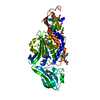
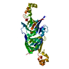
- PDB-2r8a: Crystal structure of the long-chain fatty acid transporter FadL m... -
+ Open data

- Basic information
| Entry | Database: PDB / ID: 2r8a | ||||||
|---|---|---|---|---|---|---|---|
| Title | Crystal structure of the long-chain fatty acid transporter FadL mutant delta N8 | ||||||
 Components Components | Long-chain fatty acid transport protein | ||||||
 Keywords Keywords | TRANSPORT PROTEIN / beta barrel / outer membrane protein / Lipid transport / Phage recognition / Transmembrane / Transport | ||||||
| Function / homology |  Function and homology information Function and homology informationlong-chain fatty acid transporting porin activity / ligand-gated channel activity / long-chain fatty acid transport / cell outer membrane Similarity search - Function | ||||||
| Biological species |  | ||||||
| Method |  X-RAY DIFFRACTION / SYNCHROTRON / MOLECULAR REPLACEMENT / Resolution: 3 Å X-RAY DIFFRACTION / SYNCHROTRON / MOLECULAR REPLACEMENT / Resolution: 3 Å | ||||||
 Authors Authors | Hearn, E.M. / Patel, D.R. / Lepore, B.W. / Indic, M. / van den Berg, B. | ||||||
 Citation Citation | Journal: Proc.Natl.Acad.Sci.USA / Year: 2011 Title: From the Cover: Ligand-gated diffusion across the bacterial outer membrane. Authors: Lepore, B.W. / Indic, M. / Pham, H. / Hearn, E.M. / Patel, D.R. / van den Berg, B. #1: Journal: Science / Year: 2004Title: Crystal structure of the long-chain fatty acid transporter FadL Authors: van den Berg, B. / Black, P.N. / Clemons Jr., W.M. / Rapoport, T.A. | ||||||
| History |
|
- Structure visualization
| Structure viewer | Molecule: MolmilJmol/JSmol |
|---|
- Downloads & links
-Download
| PDBx/mmCIF format | 2r8a.cif.gz | 152 KB | Display | PDBx/mmCIF format |
|---|---|---|---|---|
| PDB format | pdb2r8a.ent.gz | 119 KB | Display | PDB format |
| PDBx/mmJSON format | 2r8a.json.gz | Tree view | PDBx/mmJSON format | |
| Others |  Other downloads Other downloads |
-Validation report
| Arichive directory | https://data.pdbj.org/pub/pdb/validation_reports/r8/2r8aftp://data.pdbj.org/pub/pdb/validation_reports/r8/2r8a | HTTPS FTP |
|---|
-Related structure data
| Related structure data |  2r89C  3pf1C  3pgrC  3pgsC  3pguC  1t16S  2r4m S: Starting model for refinement C: citing same article ( |
|---|---|
| Similar structure data |


-Links
 PDBj
PDBj- Assembly
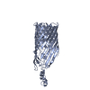
| Deposited unit | 
| ||||||||
|---|---|---|---|---|---|---|---|---|---|
| 1 | 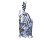
| ||||||||
| 2 | 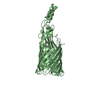
| ||||||||
| Unit cell |
|
-Components
| #1: Protein | Mass: 45801.949 Da / Num. of mol.: 2 Source method: isolated from a genetically manipulated source Source: (gene. exp.) #2: Chemical | ChemComp-C8E / (   Mass: 306.438 Da / Num. of mol.: 4 / Source method: obtained synthetically / Formula: C16H34O5 / Comment: C8E, detergent*YM Mass: 306.438 Da / Num. of mol.: 4 / Source method: obtained synthetically / Formula: C16H34O5 / Comment: C8E, detergent*YM |
|---|
-Experimental details
-Experiment
| Experiment | Method: X-RAY DIFFRACTION / Number of used crystals: 1 |
|---|
- Sample preparation
| Crystal | Density Matthews: 3.15 Å3/Da / Density % sol: 60.94 % |
|---|---|
| Crystal grow | Temperature: 295 K / Method: vapor diffusion, hanging drop / pH: 7.5 Details: 20% PEG 10000, 8% ethylene glycol, 0.1M Hepes, pH 7.5, VAPOR DIFFUSION, HANGING DROP, temperature 295K |
-Data collection
| Diffraction | Mean temperature: 100 K |
|---|---|
| Diffraction source | Source: SYNCHROTRON / Site: NSLS  / Beamline: X6A / Wavelength: 1.1 Å / Beamline: X6A / Wavelength: 1.1 Å |
| Detector | Type: ADSC QUANTUM 210 / Detector: CCD / Date: Oct 6, 2005 |
| Radiation | Monochromator: Si(111) channel cut monochromator / Protocol: SINGLE WAVELENGTH / Monochromatic (M) / Laue (L): M / Scattering type: x-ray |
| Radiation wavelength | Wavelength: 1.1 Å / Relative weight: 1 |
| Reflection | Resolution: 3→37.93 Å / Num. all: 17938 / Num. obs: 17938 / % possible obs: 80.6 % / Observed criterion σ(F): 0 / Observed criterion σ(I): 2 / Redundancy: 4.1 % / Rmerge(I) obs: 0.052 / Net I/σ(I): 13.9 |
| Reflection shell | Resolution: 3→3.11 Å / Redundancy: 3.7 % / Rmerge(I) obs: 0.511 / Mean I/σ(I) obs: 2.9 / Num. unique all: 1514 / Rsym value: 0.511 / % possible all: 68.3 |
- Processing
| Software |
| |||||||||||||||||||||||||||||||||||||||||||||||||||||||||||||||
|---|---|---|---|---|---|---|---|---|---|---|---|---|---|---|---|---|---|---|---|---|---|---|---|---|---|---|---|---|---|---|---|---|---|---|---|---|---|---|---|---|---|---|---|---|---|---|---|---|---|---|---|---|---|---|---|---|---|---|---|---|---|---|---|---|
| Refinement | Method to determine structure: MOLECULAR REPLACEMENT Starting model: PDB ENTRY 1T16 Resolution: 3→10 Å / Cross valid method: THROUGHOUT / σ(F): 0 / σ(I): 0 / Stereochemistry target values: Engh & Huber
| |||||||||||||||||||||||||||||||||||||||||||||||||||||||||||||||
| Displacement parameters | Biso mean: 46.69 Å2
| |||||||||||||||||||||||||||||||||||||||||||||||||||||||||||||||
| Refine analyze |
| |||||||||||||||||||||||||||||||||||||||||||||||||||||||||||||||
| Refinement step | Cycle: LAST / Resolution: 3→10 Å
| |||||||||||||||||||||||||||||||||||||||||||||||||||||||||||||||
| Refine LS restraints |
| |||||||||||||||||||||||||||||||||||||||||||||||||||||||||||||||
| LS refinement shell | Refine-ID: X-RAY DIFFRACTION
|
