 Movie
Movie Controller

[English] 日本語
 Yorodumi
- PDB-2r88: Crystal structure of the long-chain fatty acid transporter FadL m... -
+ Open data

- Basic information
| Entry | Database: PDB / ID: 2r88 | ||||||
|---|---|---|---|---|---|---|---|
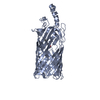
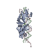
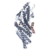
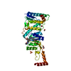
| Title | Crystal structure of the long-chain fatty acid transporter FadL mutant delta S3 kink | ||||||
 Components Components | Long-chain fatty acid transport protein | ||||||
 Keywords Keywords | TRANSPORT PROTEIN / beta barrel / outer membrane protein / Lipid transport / Phage recognition / Transmembrane / Transport | ||||||
| Function / homology |  Function and homology information Function and homology informationlong-chain fatty acid transporting porin activity / ligand-gated channel activity / long-chain fatty acid transport / cell outer membrane Similarity search - Function | ||||||
| Biological species |  | ||||||
| Method |  X-RAY DIFFRACTION / SYNCHROTRON / MOLECULAR REPLACEMENT / Resolution: 2.6 Å X-RAY DIFFRACTION / SYNCHROTRON / MOLECULAR REPLACEMENT / Resolution: 2.6 Å | ||||||
 Authors Authors | Hearn, E.M. / Patel, D.R. / Lepore, B.W. / Indic, M. / van den Berg, B. | ||||||
 Citation Citation | Journal: Nature / Year: 2009 Title: Transmembrane passage of hydrophobic compounds through a protein channel wall Authors: Hearn, E.M. / Patel, D.R. / Lepore, B.W. / Indic, M. / van den Berg, B. #1: Journal: Science / Year: 2004Title: Crystal structure of the long-chain fatty acid transporter FadL Authors: van den Berg, B. / Black, P.N. / Clemons Jr., W.M. / Rapoport, T.A. | ||||||
| History |
|
- Structure visualization
| Structure viewer | Molecule: MolmilJmol/JSmol |
|---|
- Downloads & links
-Download
| PDBx/mmCIF format | 2r88.cif.gz | 148.6 KB | Display | PDBx/mmCIF format |
|---|---|---|---|---|
| PDB format | pdb2r88.ent.gz | 117.6 KB | Display | PDB format |
| PDBx/mmJSON format | 2r88.json.gz | Tree view | PDBx/mmJSON format | |
| Others |  Other downloads Other downloads |
-Validation report
| Arichive directory | https://data.pdbj.org/pub/pdb/validation_reports/r8/2r88ftp://data.pdbj.org/pub/pdb/validation_reports/r8/2r88 | HTTPS FTP |
|---|
-Related structure data
| Related structure data | 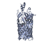 2r4lC 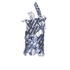 2r4nC 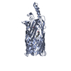 2r4oC  2r4pC  3dwnC  3dwoC  1t16S  2r4m S: Starting model for refinement C: citing same article ( |
|---|---|
| Similar structure data |


-Links
 PDBj
PDBj- Assembly
| Deposited unit | 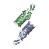
| ||||||||
|---|---|---|---|---|---|---|---|---|---|
| 1 | 
| ||||||||
| 2 | 
| ||||||||
| Unit cell |
|
-Components
| #1: Protein | Mass: 46633.801 Da / Num. of mol.: 2 Source method: isolated from a genetically manipulated source Source: (gene. exp.) Sequence details | SEQUENCE RESIDUES S100, N101, Y102, AND G103 WERE REPLACED WITH RESIDUES ALA, ASN, AND ASP TO FORM ...SEQUENCE RESIDUES S100, N101, Y102, AND G103 WERE REPLACED WITH RESIDUES ALA, ASN, AND ASP TO FORM THE DELTA S3 KINK. | |
|---|
-Experimental details
-Experiment
| Experiment | Method: X-RAY DIFFRACTION / Number of used crystals: 1 |
|---|
- Sample preparation
| Crystal | Density Matthews: 3.04 Å3/Da / Density % sol: 59.58 % |
|---|---|
| Crystal grow | Temperature: 295 K / Method: vapor diffusion, hanging drop / pH: 6.5 Details: 30% PEG 5000 MME, 0.2M ammonium sulfate, 0.1M MES, pH 6.5, VAPOR DIFFUSION, HANGING DROP, temperature 295K |
-Data collection
| Diffraction | Mean temperature: 100 K |
|---|---|
| Diffraction source | Source: SYNCHROTRON / Site: NSLS  / Beamline: X6A / Wavelength: 1.1 Å / Beamline: X6A / Wavelength: 1.1 Å |
| Detector | Type: ADSC QUANTUM 210 / Detector: CCD / Date: Apr 22, 2007 |
| Radiation | Monochromator: Si(111) channel cut monochromator / Protocol: SINGLE WAVELENGTH / Monochromatic (M) / Laue (L): M / Scattering type: x-ray |
| Radiation wavelength | Wavelength: 1.1 Å / Relative weight: 1 |
| Reflection | Resolution: 2.55→31.9 Å / Num. obs: 33965 / % possible obs: 91.9 % / Observed criterion σ(F): 0 / Observed criterion σ(I): 2.3 / Redundancy: 8.9 % / Biso Wilson estimate: 47.5 Å2 / Rmerge(I) obs: 0.079 / Net I/σ(I): 13.9 |
| Reflection shell | Resolution: 2.55→2.64 Å / Redundancy: 4.5 % / Rmerge(I) obs: 0.563 / Mean I/σ(I) obs: 2.4 / Num. unique all: 3041 / Rsym value: 0.563 / % possible all: 84.2 |
- Processing
| Software |
| |||||||||||||||||||||||||||||||||||||||||||||||||||||||||||||||
|---|---|---|---|---|---|---|---|---|---|---|---|---|---|---|---|---|---|---|---|---|---|---|---|---|---|---|---|---|---|---|---|---|---|---|---|---|---|---|---|---|---|---|---|---|---|---|---|---|---|---|---|---|---|---|---|---|---|---|---|---|---|---|---|---|
| Refinement | Method to determine structure: MOLECULAR REPLACEMENT Starting model: PDB ENTRY 1T16 Resolution: 2.6→10 Å / Cross valid method: THROUGHOUT / σ(F): 0 / σ(I): 0 / Stereochemistry target values: Engh & Huber
| |||||||||||||||||||||||||||||||||||||||||||||||||||||||||||||||
| Displacement parameters | Biso mean: 49.67 Å2
| |||||||||||||||||||||||||||||||||||||||||||||||||||||||||||||||
| Refine analyze |
| |||||||||||||||||||||||||||||||||||||||||||||||||||||||||||||||
| Refinement step | Cycle: LAST / Resolution: 2.6→10 Å
| |||||||||||||||||||||||||||||||||||||||||||||||||||||||||||||||
| Refine LS restraints |
| |||||||||||||||||||||||||||||||||||||||||||||||||||||||||||||||
| LS refinement shell | Refine-ID: X-RAY DIFFRACTION
|
