 Movie
Movie Controller


+ Open data

- Basic information
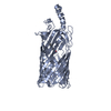
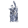
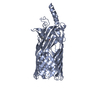
| Entry | Database: PDB / ID: 3pf1 | ||||||
|---|---|---|---|---|---|---|---|
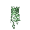
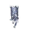
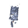
| Title | E. coli FadL Asp348Ala mutant | ||||||
 Components Components | Long-chain fatty acid transport protein | ||||||
 Keywords Keywords | LIPID TRANSPORT / outer membrane protein / oleate / oleic / beta barrel | ||||||
| Function / homology |  Function and homology information Function and homology informationlong-chain fatty acid transporting porin activity / ligand-gated channel activity / long-chain fatty acid transport / cell outer membrane Similarity search - Function | ||||||
| Biological species |  | ||||||
| Method |  X-RAY DIFFRACTION / SYNCHROTRON / MOLECULAR REPLACEMENT / Resolution: 2.7 Å X-RAY DIFFRACTION / SYNCHROTRON / MOLECULAR REPLACEMENT / Resolution: 2.7 Å | ||||||
 Authors Authors | Vandenberg, B. / Lepore, B.W. / Hearn, E.M. / Indic, M. / Patel, D. | ||||||
 Citation Citation | Journal: Proc.Natl.Acad.Sci.USA / Year: 2011 Title: From the Cover: Ligand-gated diffusion across the bacterial outer membrane. Authors: Lepore, B.W. / Indic, M. / Pham, H. / Hearn, E.M. / Patel, D.R. / van den Berg, B. | ||||||
| History |
|
- Structure visualization
| Structure viewer | Molecule: MolmilJmol/JSmol |
|---|
- Downloads & links
-Download
| PDBx/mmCIF format | 3pf1.cif.gz | 335.6 KB | Display | PDBx/mmCIF format |
|---|---|---|---|---|
| PDB format | pdb3pf1.ent.gz | 275.2 KB | Display | PDB format |
| PDBx/mmJSON format | 3pf1.json.gz | Tree view | PDBx/mmJSON format | |
| Others |  Other downloads Other downloads |
-Validation report
| Arichive directory | https://data.pdbj.org/pub/pdb/validation_reports/pf/3pf1ftp://data.pdbj.org/pub/pdb/validation_reports/pf/3pf1 | HTTPS FTP |
|---|
-Related structure data
| Related structure data |  2r89C  2r8aC  3pgrC 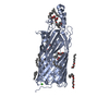 3pgsC  3pguC  1t16S S: Starting model for refinement C: citing same article ( |
|---|---|
| Similar structure data |


-Links
 PDBj
PDBj- Assembly
| Deposited unit | 
| ||||||||||||||||||
|---|---|---|---|---|---|---|---|---|---|---|---|---|---|---|---|---|---|---|---|
| 1 | 
| ||||||||||||||||||
| 2 | 
| ||||||||||||||||||
| Unit cell |
| ||||||||||||||||||
| Noncrystallographic symmetry (NCS) | NCS domain:
NCS domain segments:
|
-Components
| #1: Protein | Mass: 46308.555 Da / Num. of mol.: 2 / Fragment: mature form (UNP residues 26-446) / Mutation: D348A Source method: isolated from a genetically manipulated source Source: (gene. exp.) #2: Chemical | ChemComp-C8E / (   Mass: 306.438 Da / Num. of mol.: 8 / Source method: obtained synthetically / Formula: C16H34O5 / Comment: C8E, detergent*YM Mass: 306.438 Da / Num. of mol.: 8 / Source method: obtained synthetically / Formula: C16H34O5 / Comment: C8E, detergent*YM#3: Chemical |   Mass: 229.402 Da / Num. of mol.: 3 / Source method: obtained synthetically / Formula: C14H31NO / Comment: LDAO, detergent*YM Mass: 229.402 Da / Num. of mol.: 3 / Source method: obtained synthetically / Formula: C14H31NO / Comment: LDAO, detergent*YM#4: Water | ChemComp-HOH / |  Mass: 18.015 Da / Num. of mol.: 90 / Source method: isolated from a natural source / Formula: H2O Mass: 18.015 Da / Num. of mol.: 90 / Source method: isolated from a natural source / Formula: H2O |
|---|
-Experimental details
-Experiment
| Experiment | Method: X-RAY DIFFRACTION / Number of used crystals: 1 |
|---|
- Sample preparation
| Crystal | Density Matthews: 3.75 Å3/Da / Density % sol: 67.22 % |
|---|---|
| Crystal grow | Temperature: 295 K / Method: vapor diffusion, hanging drop / pH: 6.3 Details: 15-17% PEG 4K 0.2M KCl, protein dialysed in 10mM NaOAc 50mM NaCl, 10% glycerol 0.4%C8E4, pH 5.5, VAPOR DIFFUSION, HANGING DROP, temperature 295K |
-Data collection
| Diffraction | Mean temperature: 100 K |
|---|---|
| Diffraction source | Source: SYNCHROTRON / Site: NSLS  / Beamline: X6A / Wavelength: 0.97981 Å / Beamline: X6A / Wavelength: 0.97981 Å |
| Detector | Type: ADSC QUANTUM 4 / Detector: CCD / Date: Jun 1, 2008 / Details: toroidal mirror |
| Radiation | Monochromator: Si(111) channel cut monochromator / Protocol: SINGLE WAVELENGTH / Monochromatic (M) / Laue (L): M / Scattering type: x-ray |
| Radiation wavelength | Wavelength: 0.97981 Å / Relative weight: 1 |
| Reflection | Resolution: 2.7→47 Å / Num. all: 39050 / Num. obs: 39050 / % possible obs: 98.4 % / Observed criterion σ(F): 0 / Observed criterion σ(I): -3 / Redundancy: 4.6 % / Rmerge(I) obs: 0.118 / Rsym value: 0.118 / Net I/σ(I): 11.4 |
| Reflection shell | Resolution: 2.7→2.95 Å / Redundancy: 4.4 % / Rmerge(I) obs: 0.514 / Mean I/σ(I) obs: 4 / Rsym value: 0.514 / % possible all: 98.4 |
- Processing
| Software |
| |||||||||||||||||||||||||||||||||||||||||||||||||||||||||||||||||||||||||||||||||||||||||||||||||||||||||
|---|---|---|---|---|---|---|---|---|---|---|---|---|---|---|---|---|---|---|---|---|---|---|---|---|---|---|---|---|---|---|---|---|---|---|---|---|---|---|---|---|---|---|---|---|---|---|---|---|---|---|---|---|---|---|---|---|---|---|---|---|---|---|---|---|---|---|---|---|---|---|---|---|---|---|---|---|---|---|---|---|---|---|---|---|---|---|---|---|---|---|---|---|---|---|---|---|---|---|---|---|---|---|---|---|---|---|
| Refinement | Method to determine structure: MOLECULAR REPLACEMENT Starting model: PDB ENTRY 1T16 Resolution: 2.7→41.48 Å / SU ML: 1.06 Isotropic thermal model: TLS-derived anisotropic protein, isotropic ligands and solvent σ(F): 1.34 / Phase error: 25.85 / Stereochemistry target values: ML Details: RMS average Biso for solvent/ligands. Anisotropic scale factor is reported as Anisotropic B
| |||||||||||||||||||||||||||||||||||||||||||||||||||||||||||||||||||||||||||||||||||||||||||||||||||||||||
| Solvent computation | Shrinkage radii: 0.72 Å / VDW probe radii: 1 Å / Solvent model: FLAT BULK SOLVENT MODEL / Bsol: 38.676 Å2 / ksol: 0.4 e/Å3 | |||||||||||||||||||||||||||||||||||||||||||||||||||||||||||||||||||||||||||||||||||||||||||||||||||||||||
| Displacement parameters |
| |||||||||||||||||||||||||||||||||||||||||||||||||||||||||||||||||||||||||||||||||||||||||||||||||||||||||
| Refinement step | Cycle: LAST / Resolution: 2.7→41.48 Å
| |||||||||||||||||||||||||||||||||||||||||||||||||||||||||||||||||||||||||||||||||||||||||||||||||||||||||
| Refine LS restraints |
| |||||||||||||||||||||||||||||||||||||||||||||||||||||||||||||||||||||||||||||||||||||||||||||||||||||||||
| Refine LS restraints NCS |
| |||||||||||||||||||||||||||||||||||||||||||||||||||||||||||||||||||||||||||||||||||||||||||||||||||||||||
| LS refinement shell |
| |||||||||||||||||||||||||||||||||||||||||||||||||||||||||||||||||||||||||||||||||||||||||||||||||||||||||
| Refinement TLS params. | Method: refined / Refine-ID: X-RAY DIFFRACTION
| |||||||||||||||||||||||||||||||||||||||||||||||||||||||||||||||||||||||||||||||||||||||||||||||||||||||||
| Refinement TLS group |
|
