 Movie
Movie Controller

[English] 日本語
 Yorodumi
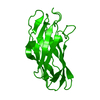
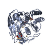
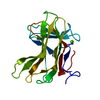
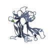
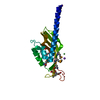
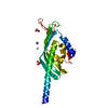
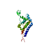
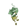
- PDB-2ccl: THE S45A, T46A MUTANT OF THE TYPE I COHESIN-DOCKERIN COMPLEX FROM... -
+ Open data

- Basic information
| Entry | Database: PDB / ID: 2ccl | ||||||
|---|---|---|---|---|---|---|---|
| Title | THE S45A, T46A MUTANT OF THE TYPE I COHESIN-DOCKERIN COMPLEX FROM THE CELLULOSOME OF CLOSTRIDIUM THERMOCELLUM | ||||||
 Components Components |
| ||||||
 Keywords Keywords | CELL ADHESION / COHESIN-DOCKERIN COMPLEX / CELLULOSOME / COHESIN / DOCKERIN / CLOSTRIDIUM THERMOCELLUM / SCAFFOLDING / CELLULOSE DEGRADATION / HYDROLASE / GLYCOSIDASE | ||||||
| Function / homology |  Function and homology information Function and homology informationcellulosome / cellulose binding / endo-1,4-beta-xylanase / endo-1,4-beta-xylanase activity / xylan catabolic process / hydrolase activity, hydrolyzing O-glycosyl compounds / cellulose catabolic process / cell wall organization / extracellular region Similarity search - Function | ||||||
| Biological species |  CLOSTRIDIUM THERMOCELLUM (bacteria) CLOSTRIDIUM THERMOCELLUM (bacteria) | ||||||
| Method |  X-RAY DIFFRACTION / SYNCHROTRON / MOLECULAR REPLACEMENT / Resolution: 2.03 Å X-RAY DIFFRACTION / SYNCHROTRON / MOLECULAR REPLACEMENT / Resolution: 2.03 Å | ||||||
 Authors Authors | Carvalho, A.L. / Dias, F.M.V. / Prates, J.A.M. / Ferreira, L.M.A. / Gilbert, H.J. / Davies, G.J. / Romao, M.J. / Fontes, C.M.G.A. | ||||||
 Citation Citation | Journal: Proc.Natl.Acad.Sci.USA / Year: 2007 Title: Evidence for a Dual Binding Mode of Dockerin Modules to Cohesins. Authors: Carvalho, A.L. / Dias, F.M.V. / Nagy, T. / Prates, J.A.M. / Proctor, M.R. / Smith, N. / Bayer, E.A. / Davies, G.J. / Ferreira, L.M.A. / Romao, M.J. / Fontes, C.M.G.A. / Gilbert, H.J. | ||||||
| History |
|
- Structure visualization
| Structure viewer | Molecule: MolmilJmol/JSmol |
|---|
- Downloads & links
-Download
| PDBx/mmCIF format | 2ccl.cif.gz | 104.7 KB | Display | PDBx/mmCIF format |
|---|---|---|---|---|
| PDB format | pdb2ccl.ent.gz | 79.6 KB | Display | PDB format |
| PDBx/mmJSON format | 2ccl.json.gz | Tree view | PDBx/mmJSON format | |
| Others |  Other downloads Other downloads |
-Validation report
| Arichive directory | https://data.pdbj.org/pub/pdb/validation_reports/cc/2cclftp://data.pdbj.org/pub/pdb/validation_reports/cc/2ccl | HTTPS FTP |
|---|
-Related structure data
| Related structure data |  1ohzS S: Starting model for refinement |
|---|---|
| Similar structure data |


-Links
 PDBj
PDBj

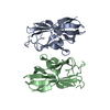
- Assembly
| Deposited unit | 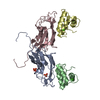
| ||||||||
|---|---|---|---|---|---|---|---|---|---|
| 1 | 
| ||||||||
| 2 | 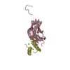
| ||||||||
| Unit cell |
|
-Components
| #1: Protein | Mass: 16956.029 Da / Num. of mol.: 2 / Fragment: COHESIN 2 DOMAIN, RESIDUES 181-328 Source method: isolated from a genetically manipulated source Source: (gene. exp.) CLOSTRIDIUM THERMOCELLUM (bacteria) / Production host: #2: Protein | Mass: 6794.856 Da / Num. of mol.: 2 / Fragment: RESIDUES 730-791 / Mutation: YES Source method: isolated from a genetically manipulated source Source: (gene. exp.) CLOSTRIDIUM THERMOCELLUM (bacteria) / Production host: #3: Chemical |   Mass: 94.971 Da / Num. of mol.: 2 / Source method: obtained synthetically / Formula: PO4 Mass: 94.971 Da / Num. of mol.: 2 / Source method: obtained synthetically / Formula: PO4#4: Chemical | ChemComp-CA /   Mass: 40.078 Da / Num. of mol.: 4 / Source method: obtained synthetically / Formula: Ca Mass: 40.078 Da / Num. of mol.: 4 / Source method: obtained synthetically / Formula: Ca#5: Water | ChemComp-HOH / |  Mass: 18.015 Da / Num. of mol.: 496 / Source method: isolated from a natural source / Formula: H2O Mass: 18.015 Da / Num. of mol.: 496 / Source method: isolated from a natural source / Formula: H2OCompound details | ENGINEERED RESIDUE IN CHAIN B, SER 777 TO ALA ENGINEERED RESIDUE IN CHAIN B, THR 778 TO ALA ...ENGINEERED | |
|---|
-Experimental details
-Experiment
| Experiment | Method: X-RAY DIFFRACTION |
|---|
- Sample preparation
| Crystal | Density Matthews: 2.2 Å3/Da / Density % sol: 42.4 % |
|---|
-Data collection
| Diffraction | Mean temperature: 100 K |
|---|---|
| Diffraction source | Source: SYNCHROTRON / Site: ESRF  / Beamline: ID14-1 / Wavelength: 0.931 / Beamline: ID14-1 / Wavelength: 0.931 |
| Detector | Type: ADSC CCD / Detector: CCD |
| Radiation | Protocol: SINGLE WAVELENGTH / Monochromatic (M) / Laue (L): M / Scattering type: x-ray |
| Radiation wavelength | Wavelength: 0.931 Å / Relative weight: 1 |
| Reflection | Resolution: 2→28.41 Å / Num. obs: 227515 / % possible obs: 95.8 % / Observed criterion σ(I): 2.8 / Redundancy: 3.6 % / Rmerge(I) obs: 0.1 / Net I/σ(I): 7.1 |
| Reflection shell | Resolution: 2→2.11 Å / Redundancy: 3.3 % / Rmerge(I) obs: 0.02 / Mean I/σ(I) obs: 29.1 / % possible all: 95.8 |
- Processing
| Software |
| ||||||||||||||||||||||||||||||||||||||||||||||||||||||||||||||||||||||||||||||||||||||||||||||||||||||||||||||||||||||||||||||||||||||||||||||||||||||||||||||||||||||||||||||||||||||
|---|---|---|---|---|---|---|---|---|---|---|---|---|---|---|---|---|---|---|---|---|---|---|---|---|---|---|---|---|---|---|---|---|---|---|---|---|---|---|---|---|---|---|---|---|---|---|---|---|---|---|---|---|---|---|---|---|---|---|---|---|---|---|---|---|---|---|---|---|---|---|---|---|---|---|---|---|---|---|---|---|---|---|---|---|---|---|---|---|---|---|---|---|---|---|---|---|---|---|---|---|---|---|---|---|---|---|---|---|---|---|---|---|---|---|---|---|---|---|---|---|---|---|---|---|---|---|---|---|---|---|---|---|---|---|---|---|---|---|---|---|---|---|---|---|---|---|---|---|---|---|---|---|---|---|---|---|---|---|---|---|---|---|---|---|---|---|---|---|---|---|---|---|---|---|---|---|---|---|---|---|---|---|---|
| Refinement | Method to determine structure: MOLECULAR REPLACEMENT Starting model: PDB ENTRY 1OHZ Resolution: 2.03→20 Å / Cor.coef. Fo:Fc: 0.931 / Cor.coef. Fo:Fc free: 0.887 / SU B: 7.266 / SU ML: 0.115 / TLS residual ADP flag: LIKELY RESIDUAL / Cross valid method: THROUGHOUT / ESU R: 0.221 / ESU R Free: 0.185 / Stereochemistry target values: MAXIMUM LIKELIHOOD / Details: HYDROGENS HAVE BEEN ADDED IN THE RIDING POSITIONS.
| ||||||||||||||||||||||||||||||||||||||||||||||||||||||||||||||||||||||||||||||||||||||||||||||||||||||||||||||||||||||||||||||||||||||||||||||||||||||||||||||||||||||||||||||||||||||
| Solvent computation | Ion probe radii: 0.8 Å / Shrinkage radii: 0.8 Å / VDW probe radii: 1.4 Å / Solvent model: MASK | ||||||||||||||||||||||||||||||||||||||||||||||||||||||||||||||||||||||||||||||||||||||||||||||||||||||||||||||||||||||||||||||||||||||||||||||||||||||||||||||||||||||||||||||||||||||
| Displacement parameters | Biso mean: 10.2 Å2
| ||||||||||||||||||||||||||||||||||||||||||||||||||||||||||||||||||||||||||||||||||||||||||||||||||||||||||||||||||||||||||||||||||||||||||||||||||||||||||||||||||||||||||||||||||||||
| Refinement step | Cycle: LAST / Resolution: 2.03→20 Å
| ||||||||||||||||||||||||||||||||||||||||||||||||||||||||||||||||||||||||||||||||||||||||||||||||||||||||||||||||||||||||||||||||||||||||||||||||||||||||||||||||||||||||||||||||||||||
| Refine LS restraints |
|